9808 Lecture Notes - Lecture 2: Central Nervous System, Corpus Callosum, Dura Mater

19 Jun 2018
School
Department
Course
Professor

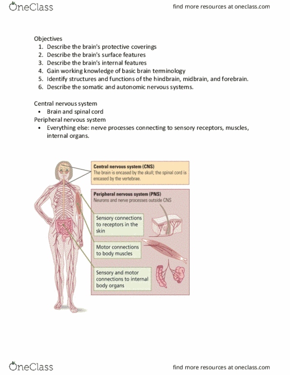
Central Nervous System – Lecture
1. Describe brain development in an embryo *** NOT TESTED ON
2. Describe the gross anatomical structures of the brain - meninges - corpus callosum -
ventricles (CSF) - cerebellum - lobes - hypothalamus
3. Describe the function of cerebrospinal fluid
4. Label the blood supply to the brain
5. Locate and describe the function of the sensory cortex
6. Locate and describe the function of the motor cortex
7. Label the internal and external anatomy of the spinal cord
8. Describe the function of each of the 12 cranial nerves
1- Brain Development in Embryo
NOT TESTED ON ***
find more resources at oneclass.com
find more resources at oneclass.com

2- Describe the gross anatomical
structures of the brain
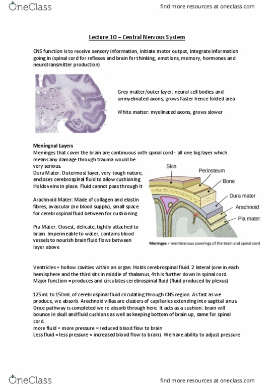
Meninges = membranous
coverings of the brain and spinal
cord
3meninges:
-dura mater - directly attached to skill, very tough / leathery, sits just under skull, made
of dense irregular tissue , main job = close cerebral spinal fluid – so it doesn’t diffuse
anywhere else and stays inside . Also holds sinuses – which are veins that drain skull and
brain.
- Arachnoid - spider like, next layer down, really fine tissue paper, composed of loosely
arranged collagen and elastin fibres.
- Pia mater bottom membranous covering, so closely attached to brain can only see with
a microscope, imperial to fluid, keeps fluid in, has blood vessels that run through
membrane into brain so we can feed the brain.
- are 3 different textures to give varying levels of protection
Menagitous = inflammation of meninges.
Corpus callosum - ventricles (CSF) - cerebellum - lobes - hypothalamus
Ventricles
- Have 4 within the brain
find more resources at oneclass.com
find more resources at oneclass.com

- Are just hollow cavity’s - in brain are filled with fluid
1&2 ventricles = lateral ventricle s= 2 c shaped, are continuous into 3rd ventricle
3rd ventricle = part in middle – fluid
flows from lateral in the 3rd
4th ventricle – connected to 3rd ventricle
by tube (cerebral aqueduct
Functions = to transport cerebral
spinal fluid, production of cerebral
spinal fluid and to remove from
system when we need to.
Functions of Cerebrospinal Fluid (CSF) =
the fluid that covers the brain, what the brain sits in
1. Protection – shock absorption, enclosed within meninges.
2. Buoyancy – reduce the weight of the brain, helps the brain float.
3. Chemical Stability – remove waste products. take nutrients to brain
4. Circulation – facilitate blood perfusion, can change how quickly it is removed from
the system, can change pressure in skull can help with blood flow.
Once has done full loop of brain and spine it gets reused.
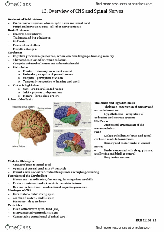
Cerebrum
Big portion at top of brain .outer region is grey matter. Inner region/ layer is white matter.
-Lumpy pits = gyri
-Fold that go in =sulci
find more resources at oneclass.com
find more resources at oneclass.com
Document Summary
Central nervous system lecture: describe brain development in an embryo *** not tested on. Describe the gross anatomical structures of the brain - meninges - corpus callosum - ventricles (csf) - cerebellum - lobes - hypothalamus. Locate and describe the function of the sensory cortex. Locate and describe the function of the motor cortex. Label the internal and external anatomy of the spinal cord. Describe the function of each of the 12 cranial nerves. 2- describe the gross anatomical structures of the brain. Meninges = membranous coverings of the brain and spinal cord. Also holds sinuses which are veins that drain skull and brain. Arachnoid - spider like, next layer down, really fine tissue paper, composed of loosely arranged collagen and elastin fibres. Corpus callosum - ventricles (csf) - cerebellum - lobes - hypothalamus. Are just hollow cavity"s - in brain are filled with fluid. 1&2 ventricles = lateral ventricle s= 2 c shaped, are continuous into 3rd ventricle.