HPE110 Lecture Notes - Lecture 6: Osteoarthritis, Anatomical Terms Of Location, Sprain


HB101-Lecture 6
Lower limb functional anatomy
-Bone morphology
-Joint degrees of freedom (DOF)
and range of motion (ROM)
-Muscular morphology
-Injury
-Functional movements
-Surface anatomy
-ROM testing
Primary support and locomotion mechanism
Humans heavily dependent due to bipedalism
Human upper and lower limb discrepancies pronounced compared to primates
Lower limb comprised of largest muscles and bones in the body
Structured to undergo the largest forces
regularly encountered by the body
Pelvis
Link between the lower limb and the trunk
Cradles the internal organs of the torso
Females wider pelvis to allow child birth
Large articular surfaces for hip and trunk muscles
Often referred to as centre of mass point in body
find more resources at oneclass.com
find more resources at oneclass.com

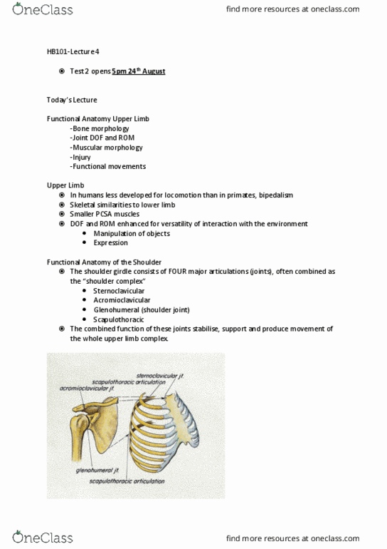
Anatomical Structure – Pelvic Girdle
Pelvic girdle
Ilium (upper)
Provides points of origin for muscles involved in hip abduction (Gmed, Gmin)
Ischium (posterior)
Origin for muscles involved in hip extension (Gmax, BF, Semiten., Seminmen.)
Pubis (anterior)
Origin for muscles involved in hip flexion (RF, Sar)
Sacrum
Left and right pelvic bones joined together posteriorly
Joints of the pelvic girdle
Pubic Symphasis
Connects left and right anterior part of the pelvis
Weak cartilaginous joint supported by a pubic ligament (linked with tendonitis –
osteitis pubis)
Sacroiliac (two – left and right)
Runs posteriorly and is the strongest joint held by powerful ligament support
(strongest in the whole human body)
Transmits body weight to the hip and subjected to lumbar loading. Also absorbs
shear forces during gait
Males have stronger joints, and thus are less flexible than females in pelvis ROM
Femoral (hip joint)
Stable, yet mobile – similar to glenohumeral joint, although reduced sub/dislocation
– strong musculature
70% of femoral head articulates with the acetabulum, compared with only 25% of
the humeral head with the glenoid cavity.
Synovial (ball & socket) joint between the femoral head and acetabulum /
acetabulum labrum (similar to glenoid fossa / labrum) at the glenohumeral joint.
find more resources at oneclass.com
find more resources at oneclass.com

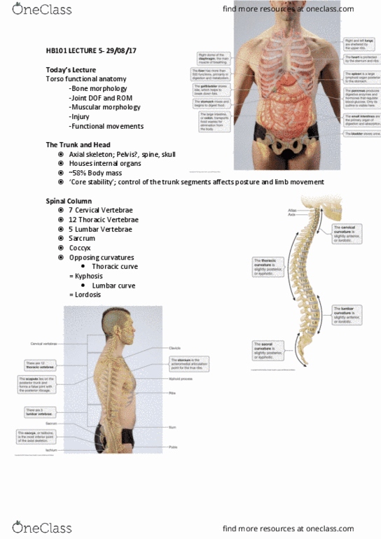
Hip Joint
Ball and socket joint; spherical femoral head (ball) and convex acetabulum (socket)
Highly mobile yet stable joint due to architecture, ligamenture and musculature
Three DOF;
flex/ext; ~120°/~15°
add/abd; ~25°/~45°
int/ext rot; ~40°/~40°
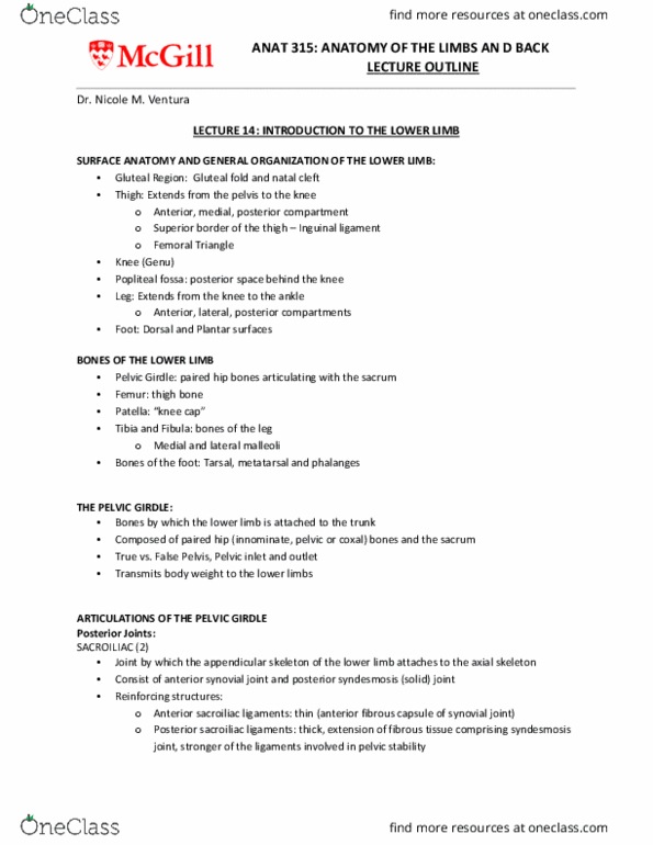
Hip & “houlder joit siilarities
Shoulder girdle
Acromioclavicular
Scapulothoracic
Sternoclavicular
Pelvic girdle
Sacroilliac
Pubic symphasis
“houlder joit
Gleohueral
Hip joit
Feoral
find more resources at oneclass.com
find more resources at oneclass.com
Document Summary
Human upper and lower limb discrepancies pronounced compared to primates. Lower limb comprised of largest muscles and bones in the body. Structured to undergo the largest forces regularly encountered by the body. Link between the lower limb and the trunk. Cradles the internal organs of the torso. Females wider pelvis to allow child birth. Large articular surfaces for hip and trunk muscles. Often referred to as centre of mass point in body. Provides points of origin for muscles involved in hip abduction (gmed, gmin) Origin for muscles involved in hip extension (gmax, bf, semiten. , seminmen. ) Origin for muscles involved in hip flexion (rf, sar) Left and right pelvic bones joined together posteriorly. Connects left and right anterior part of the pelvis. Weak cartilaginous joint supported by a pubic ligament (linked with tendonitis osteitis pubis) Runs posteriorly and is the strongest joint held by powerful ligament support (strongest in the whole human body)