BIOL 2P98 Lecture Notes - Lecture 4: Oil Immersion, Electron Microscope, Gram Staining

19 Jan 2016
School
Department
Course
Professor


Document Summary
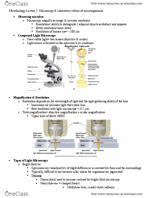
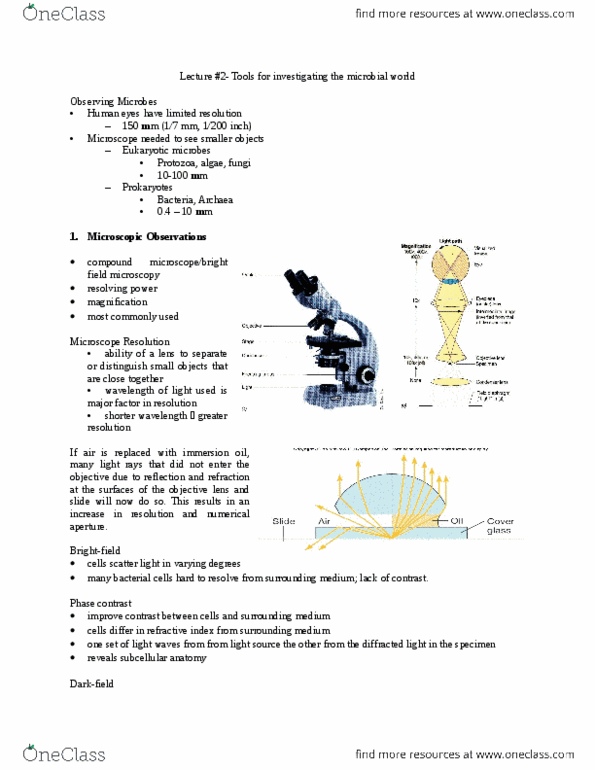
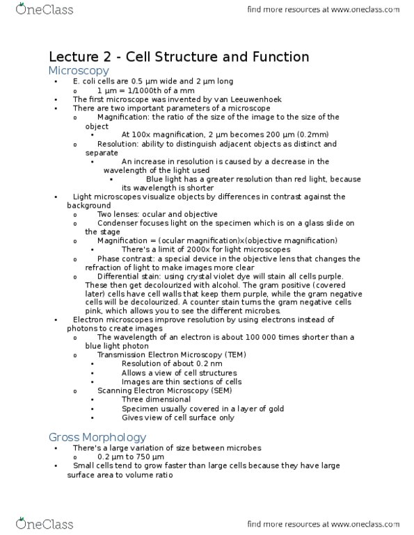
Chapters 2 & 3: microscopy & laboratory culture of. *** test dates changed: test #1 thursday october 8th. Bright-filed: specimens are visualized because of slight differences in contrast, typically difficult to see bacterial cells. Unless the organisms are between them and the surroundings pigmented. Staining: stains (dyes) can be used to increase contrast for bright-field, many dyes are positively charged (basic) microscopy, methylene blue, crystal violet, safranin, bind to negatively charged cell components (nucleic acids, many proteins, polysaccharides, cell surfaces) Differential stains: stain different kinds of cells different colours, most common: gram stain, divides baceria into two major groups: Gram-positive (purple) have thick heavy duty wall that does not allow alcohol to stain heavily. Gram-negative (pink) outer membrane with a thin cell wall and cell membrane, which allow alcohol to stain heavily (ex. E. coli: arises due to difference in cell wall structure.