BIOL 303 Lecture Notes - Lecture 8: Limb Bud, Neuromuscular Junction, Axon Terminal

14 May 2018
School
Department
Course
Professor

Biol 303 March 22nd
General
• MSURJ launch party is coming up
• MBSU elections are happening
Synapse formation and refinement
• Axons travel for a long time and eventually find their muscle target
• The nerve terminal has to specify structures to facilitate the communication between
the nerve and the muscle
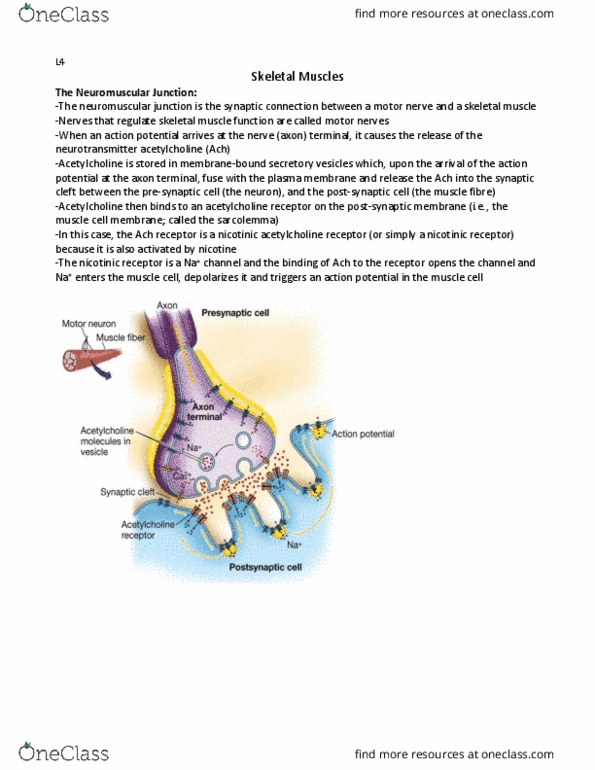
• Neuromuscular junction: considered to be a synapse between motor axon terminal and
muscle fibre
• Pre-synaptic membrane is in the motor axon/neuron → it faces the muscle membrane
AKA post-synaptic membrane
• Pre-synaptic terminal must have accumulation of synaptic vesicles
• The synaptic vesicles contain cargo of neurotransmitters
• At the neuro-muscular junction, the neurotransmitter is acetylcholine
• Post-synaptic membrane has a cluster of acetylcholine receptors
• Ach binds to Ach receptors on the muscle terminal and induces muscle contraction
Steps of communication
1. Adhesion – bringing membranes into proximity with one another
2. Neural terminal produces Agrin → a protein that induces post-synaptic development
AKA clustering of Ach receptors
3. In the muscle fibre, secreted proteins like FGF and Gdnf are produced → they can
induce presynaptic development Ex. 1) Accumulation of synaptic vesicles, 2) Clustering
of Ca2+ channels
• The end result is the accumulation of synaptic vesicles and structures that facilitates the
release of neurotransmitters AND the clustering of receptors in the receiving terminals
Synapse elimination
• In the embryo: At the junction where the motor neuron meets the muscle fibre, the
neurons are branched to have several terminals that innervate different muscle fibres
→ several neurons innervate the same muscle
• In the adult: there are less branches → only one motor neuron innervates each muscle
fibre
• This is because of synapse competition → the terminals of the different neurons
compete against each other when in the embryo and on the same muscle fibre →
whichever one has the greatest activity stays there
• Synapse competition eliminates unwanted terminals
find more resources at oneclass.com
find more resources at oneclass.com

Vertebrate Limb Development – looking at chicks
• Used as a model for organogenesis
• The early structures can be easily visualized in the developing limb with staining
• At 3 days of incubation after laying an egg → there is a small protrusion at the
wing/limb bud
• The location of the limb buds only occurs at specific locations along the anterior-
posterior axis
• What are the steps in the development?
Limb induction
• The mature limb comes from the limb bud
• How does the embryo know which region will give rise to the limb bud?
• 1) Specification of the limb field → can be identified by:
• Selective ablation: remove specific tissues and see if it affects development of the limb
• Labelling: see which tissues travel where from early to late embryo
• Transplantation: which tissues may give rise to future limb structures and can they make
them elsewhere
• What is the key specifier of limb bud location?
Hox genes
• Lots of evidence suggests that Hox genes (b9, c9, and d9) are involved because their
expression changes during limb bud formation
• Hox 4 and 5 might also be involved
• Tbx5 and Tbx 4 → function downstream of Hox genes
• The expressio of differet Hox gees speifies li field ad idue the speifi
expression of Tbx4 and 5 in their specific fields
• Wing/forelimb region expresses Tbx5
• Tbx4 is expressed in the leg/hindlimb region
• Tx ad are trasriptio fators that regulate li iduer gees dowstrea:
FGF10 → their function is to stimulate the expression FGF10 in mesoderm cells AKA
lateral plate mesoderm, which give rise to later cartilage cells that will become bone
• Many people consider FGF10 as a direct limb inducer
Experiment
• Use porous beads and soaked them with purified FGF10
• You can embed those beads, which will release FGF10, into a tissue that does not
normally produce a limb → the result is that they give rise to an ectopic wing or leg
(depending on whether you put the beads near an existing wing or leg structure)
• The type of limb bud it gives rise to will depend on where it is place → there is some
importance of location
How does the presence of FGF10 lead to limb bud formation?
find more resources at oneclass.com
find more resources at oneclass.com
Document Summary
General: msurj launch party is coming up, mbsu elections are happening. Steps of communication: adhesion bringing membranes into proximity with one another, neural terminal produces agrin a protein that induces post-synaptic development. Aka clustering of ach receptors: in the muscle fibre, secreted proteins like fgf and gdnf are produced they can induce presynaptic development ex. 1) accumulation of synaptic vesicles, 2) clustering of ca2+ channels: the end result is the accumulation of synaptic vesicles and structures that facilitates the release of neurotransmitters and the clustering of receptors in the receiving terminals. In the embryo: at the junction where the motor neuron meets the muscle fibre, the neurons are branched to have several terminals that innervate different muscle fibres. Fgf10 their function is to stimulate the expression fgf10 in mesoderm cells aka lateral plate mesoderm, which give rise to later cartilage cells that will become bone: many people consider fgf10 as a direct limb inducer.