ANAT 261 Lecture Notes - Type I Collagen

15 Mar 2012
School
Department
Course
Professor
Document Summary
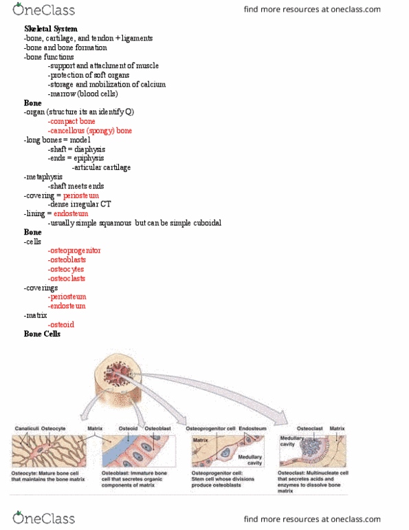
Two types of bone: trabecular (or spongy or cancellous) bone, woven (immature, disorganized fibres, where: ends of long bones, craniofacial bones, immature bone, thin bones, compact bone, lamellar (mature, organized fibres, where: shafts of long bones. Extracellular matrix is predominantly collagen type i. Endosteum: a single layer of osteoblasts and osteoprogenitor cells that line the marrow cavity inner circumferential system. Periosteum ct sheath that surrounds bone: made of two layers: Allows long bones to grow in length. At the junction of epiphysis (end) and diaphysis (shaft) from outside to inside: secondary ossification center of endochondral ossification, zone of resting cartilage. Hyaline cartilage with chondrocytes and isogenic groups: zone of proliferation rapidly dividing chondrocytes (interstitial growth) Cells look like a stack of coins: zone of hypertrophy. Cartilage ecm begins to calcify (ecm appears to fade: zone of cell death. Empty lacuna: zone of mixed spicules. Osteoblasts, osteocytes, and osteoclasts can all be spotted.