PSYC 211 Lecture Notes - Lecture 19: Visual Acuity, Peripheral Vision, Vitreous Body

16 Mar 2017
School
Department
Course
Professor
Document Summary
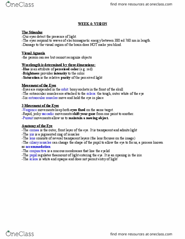
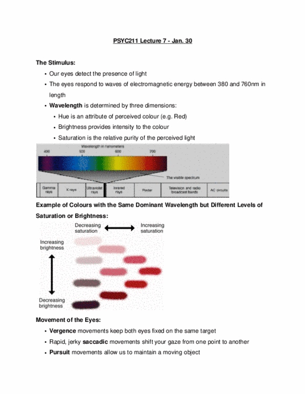
Anatomy on the eye (i) the cornea is the outer, front layer of the eye. It is transparent and admits light, sensitive to day time and night time the iris is a pigmented ring of muscle the lens consists of several transparent layers. It is an opening in the iris the sclera is opaque and does not permit entry of light. Conjunctiva is a mucous membranes that line the eye and merges with the inside if the eyelids. Anatomy of the eye (ii) the interior lining of the is the retina. It contains the highest number of cones site of blind spot is the point at which the optic nerve exists through the back of the eye. It has no receptors and therefore there is no vision. The route within the retina light passes through transparent cells and stimulate the photoreceptors located at the back of the eye.