PSYC 311 Lecture 4: Petrides The Ventrolateral Frontal Region

11 May 2018
School
Department
Course
Professor

CHAPTER
3
The Ventrolateral Frontal Region
Michael Petrides
Montreal Neurological Institute, McGill University, Montreal, Quebec, Canada
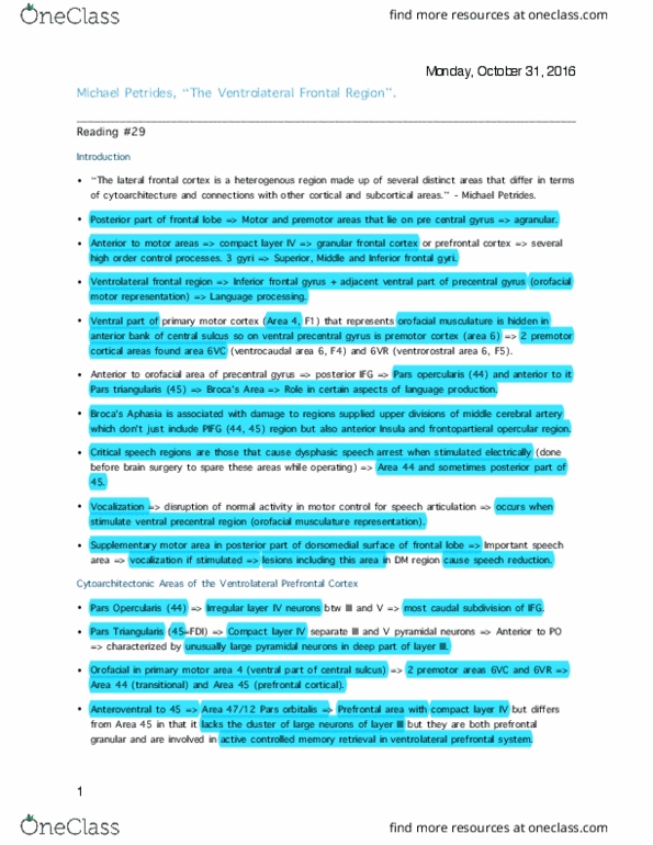
The lateral frontal cortex is a heterogeneous region
comprising several distinct areas that differ both in terms
of their cellular architecture (cytoarchitecture) and their
connections with other cortical and subcortical areas
(Figure 3.1). The posterior part of the frontal lobe
includes several motor and premotor areas that lie
mostly on the precentral gyrus (region in white in
Figure 3.1). The compact layer of small neurons (layer IV)
clearly separating the pyramids of layer III from those
of layer V in primary sensory cortex and other isocorti-
cal areas is difficult to discern in the motor/premotor
areas because these small neurons are intermixed with
larger pyramidal neurons, leading to the tradition of
referring to these areas as “agranular.” However, these
so-called agranular isocortical motor areas do have
small interneurons and must not be confused with the
truly agranular phylogenetically older areas of the
limbic region of the brain (Garcı
´a-Cabezas & Barbas,
2014). Anterior to these motor areas are several
cytoarchitectonic areas that exhibit a compact and
distinct layer IV, and this region is often referred to as
the “granular” frontal cortex or the “prefrontal” cortex.
These prefrontal cortical areas have been shown to par-
ticipate in several higher-order control processes that
regulate attention to the environment, working mem-
ory, various aspects of controlled memory retrieval,
and behavioral adjustment to changes in the environ-
ment (see reviews in Stuss & Knight, 2013). The part of
the lateral frontal cortex that extends anterior to the pre-
central gyrus is traditionally divided into three gyri: the
superior, middle, and inferior frontal gyri. The caudal
part of the prefrontal cortex on the superior and middle
frontal gyri is occupied by subdivisions of area 8, a corti-
cal region regulating attentional processes, which is
succeeded anteriorly by the mid-dorsolateral prefrontal
region (area 46 and the related areas 9/46) that plays
a major role in certain aspects of working memory,
such as the tracking of self-generated and externally
generated events in working memory (Petrides, 1996,
2013). Although we might expect the specific roles of
these areas in attentional control and working memory
to be reflected in language processing, the dorsolateral
prefrontal areas are not core language areas in the sense
that neither fundamental language comprehension nor
production is impaired.
The part of the frontal lobe that is most relevant to
language processing is the inferior frontal gyrus and
the adjacent ventral part of the precentral gyrus,
namely the ventrolateral frontal region (Figure 3.2).
The motor representation of the orofacial part of the
body is found on the ventral part of the precen-
tral gyrus (Penfield & Boldrey, 1937; Penfield &
Rasmussen, 1950). The ventral part of the primary
motor cortex (Brodmann area 4) that represents the
orofacial musculature is largely hidden in the anterior
bank of the central sulcus and, therefore, most of the
cortex on the crown of the ventral precentral gyrus is
occupied by premotor cortex (i.e., area 6) (Brodmann,
1909). More recent studies have identified two pre-
motor cortical areas on the ventral precentral gyrus, a
caudal one, area 6VC (ventrocaudal part of area 6; also
known as area F4), and a rostral area, area 6VR
(ventrorostral part of area 6; also known as area F5)
(see Petrides, 2014, for details). The terms “area F1”
(corresponding to area 4), “area F4” (corresponding to
area 6VC), and “area F5” (corresponding to area 6VR)
were proposed for comparable areas in the macaque
monkey by Matelli, Luppino, and Rizzolatti (1985).
Immediately anterior to the orofacial part of the precen-
tral gyrus lies the posterior part of the inferior frontal
gyrus, namely the pars opercularis, which is occupied
by cortical area 44 and is succeeded anteriorly by the
pars triangularis (area 45) (Figures 3.1 and 3.2).
The posterior part of the inferior frontal gyrus in the
language-dominant hemisphere is traditionally con-
sidered to be the classical Broca’s region, namely the
25Neurobiology of Language. DOI: http://dx.doi.org/10.1016/B978-0-12-407794-2.00003-1 ©2016 Elsevier Inc. All rights reserved.

frontal cortical region that plays a critical role in cer-
tain aspects of language production (Friederici, 2011;
Geschwind, 1970; Grodzinsky, 2000). Several attempts
have been made to specify more precisely the critical
zone for language within the inferior frontal gyrus on
the basis of clinicalanatomical correlation studies, but
these efforts had only limited success because lesions
in human subjects are rarely restricted to specific
subdivisions of the inferior frontal region (Mohr, 1976;
Mohr et al., 1978). The syndrome of Broca’s aphasia,
which is characterized by severe impairment in lan-
guage production (including impaired syntactic pro-
cessing), is the result of massive damage to the
territory of the upper division of the middle cerebral
artery and involves not only the cortical structures in
the posterior part of the inferior frontal gyrus (i.e.,
areas 44 and 45) but also the adjacent frontoparietal
opercular region and the anterior parts of the insula
(Ackermann & Riecker, 2004; Baldo, Wilkins, Ogar,
Willock, & Dronkers, 2011; Dronkers, 1996; Mohr,
1976). The best evidence thus far linking specific parts
of the inferior frontal gyrus to language production
has been obtained from electrical stimulation of the
cerebral cortex under local anesthesia during brain sur-
gery. In this approach that is motivated by the need to
spare cortex critical for language during brain surgery,
the critical region for speech is considered to be the
part of the cortex from which dysphasic speech arrest
can be evoked by the application of electrical
FIGURE 3.1 Cytoarchitectonic map of the lateral surface of the
human and the macaque monkey frontal lobe by Petrides and Pandya
(1994). The white region on the precentral gyrus is the primary motor
cortex (area 4) and the various subdivisions of the premotor region
(area 6). The inset shows the location of area 44 in the macaque monkey
in the fundus of the inferior limb (ramus) of the arcuate sulcus.
FIGURE 3.2 The sulcal and gyral morphology of the ventrolateral
frontal region in the human brain. The shaded region represents
the orbitofrontal cortex that is continuous with the pars orbitalis of the
inferior frontal gyrus. Abbreviations: aalf, anterior ascending ramus of
the lateral fissure (ascending sulcus, vertical sulcus); ascs, anterior
subcentral sulcus; cs, central sulcus; ds, diagonal sulcus; half, horizon-
tal anterior ramus of the lateral fissure (horizontal sulcus); IFG,
inferior frontal gyrus; ifs, inferior frontal sulcus; iprs, inferior precen-
tral sulcus; los-p, posterior ramus of the lateral orbital sulcus; MFG,
middle frontal gyrus; Op, pars opercularis of the inferior frontal
gyrus; Or, pars orbitalis of the inferior frontal gyrus; PrG, precentral
gyrus; prts, pretriangular sulcus; ScG, subcentral gyrus; STG, superior
temporal gyrus; sts, superior temporal sulcus; Tr, pars triangularis of
the inferior frontal gyrus; ts, triangular sulcus (incisura capitis).
26 3. THE VENTROLATERAL FRONTAL REGION
B. NEUROBIOLOGICAL FOUNDATIONS

stimulation (Duffau, Moritz-Gasser, & Mandonnet,
2014; Ojemann, 1992; Ojemann, Ojemann, Lettich, &
Berger, 1989; Penfield & Roberts, 1959; Rasmussen &
Milner, 1975). Dysphasic speech arrest occurs most
reliably from stimulation of the pars opercularis (area
44) (Rasmussen & Milner, 1975), although speech arrest
can also be evoked from stimulation of the posterior
part of area 45. Stimulation of the ventral precentral
region, where the orofacial musculature is represented,
also interferes with speech, primarily in the form of dys-
arthria and evoked vocalization responses caused by
disruption of normal activity in the motor circuits neces-
sary for speech articulation (Penfield & Roberts, 1959;
Rasmussen & Milner, 1975).
The studies of Penfield and colleagues established
another important region for speech on the posterior
part of the dorsomedial surface of the frontal lobe, the
supplementary motor area. Vocalization, as well as
interference with speech, can result from stimulation
of the supplementary motor area (Penfield & Welch,
1951). Several studies have shown that lesions of the
dorsomedial frontal region, which include the sup-
plementary motor area but are not restricted to it,
lead to significant reduction in speech (Chapados &
Petrides, 2013; Goldberg, 1985; Krainik et al., 2003;
Nachev, Kennard, & Husain, 2008; Rostomily, Berger,
Ojemann, & Lettich, 1991). Furthermore, three somato-
topically organized motor areas just ventral and
anterior to the supplementary motor region, originally
shown in the monkey brain (Dum & Strick, 1993), have
also been recently demonstrated in the human brain
(Amiez & Petrides, 2014), and there is some evidence
that the cingulate motor region may also play a role in
speech (Paus, Petrides, Evans, & Meyer, 1993).
3.1 CYTOARCHITECTONIC AREAS
OF THE VENTROLATERAL
PREFRONTAL CORTEX
Anterior to the ventral premotor region lies a corti-
cal area in which irregular patches of small neurons
appear between the pyramidal neurons of layers III
and V (Figure 3.3). This area that occupies the most
caudal subdivision of the inferior frontal gyrus, the
pars opercularis, is Brodmann area 44 (area FCBm of
Economo & Koskinas, 1925)(Figure 3.1). Area 44 is
succeeded anteriorly by prefrontal area 45, which lies
on the pars triangularis of the inferior frontal gyrus. In
area 45, the small neurons of layer IV create a compact
layer and, therefore, the pyramidal neurons of layers
III and V are clearly separated (compare Figures 3.3
and 3.4)(Amunts et al., 1999; Petrides & Pandya, 1994,
2002). Area 45 is further characterized by clusters of
unusually large and deeply stained pyramidal neurons
in the deep part of layer III, a characteristic that
unambiguously differentiates area 45 from the sur-
rounding prefrontal areas. This unusual characteristic of
area 45 led Economo and Koskinas (1925) to refer to it as
area FDΓ; the Greek letter Γrefers to the clusters of
giant-like neurons in layer III (Figure 3.4). In conclusion,
starting from the ventral part of the central sulcus where
the orofacial part of the primary motor cortical area 4 is
represented, and proceeding in an anterior direction,
there are two premotor areas, 6VC and 6VR, that are
succeeded by the transitional area 44 and, further
anterior, by the prefrontal cortical area 45 (Figure 3.1).
Anterior and ventral to area 45 lies area 47/12, which
occupies the pars orbitalis of the inferior frontal gyrus
(Figure 3.1). Although area 47/12 has not been tradi-
tionally considered as a core language area, recent func-
tional neuroimaging studies have suggested that it may
play a major role in the controlled access to stored con-
ceptual representations (Badre & Wagner, 2007) and
semantic unification (Zhu et al., 2012). Thus, we pro-
vide a brief discussion of its identification and cytoarch-
itecture here because the architectonic description of
FIGURE 3.3 Photomicrograph of area 44 in the human brain.
Note the interrupted layer IV, highlighted with yellow. The Roman
numerals IVI mark the six layers of the cortex. Calibration bar
equals 1 mm. From Petrides (2014) with permission from the publisher.
273.1 CYTOARCHITECTONIC AREAS OF THE VENTROLATERAL PREFRONTAL CORTEX
B. NEUROBIOLOGICAL FOUNDATIONS
Document Summary
Montreal neurological institute, mcgill university, montreal, quebec, canada. The lateral frontal cortex is a heterogeneous region comprising several distinct areas that differ both in terms of their cellular architecture (cytoarchitecture) and their connections with other cortical and subcortical areas (figure 3. 1). The posterior part of lobe includes several motor and premotor areas that lie mostly on the precentral gyrus (region in white in. Anterior to these motor areas are several cytoarchitectonic areas that exhibit a compact and distinct layer iv, and this region is often referred to as the granular frontal cortex or the prefrontal cortex. The part of the lateral frontal cortex that extends anterior to the pre- central gyrus is traditionally divided into three gyri: the superior, middle, and inferior frontal gyri. The part of the frontal lobe that is most relevant to language processing is the inferior frontal gyrus and the adjacent ventral part of the precentral gyrus, namely the ventrolateral frontal region (figure 3. 2).