PSYC 427 Lecture Notes - Lecture 3: Alpha Motor Neuron, Extraocular Muscles, Neuromuscular Junction

14 May 2018
School
Department
Course
Professor

PSYC 427 – LECTURE 3
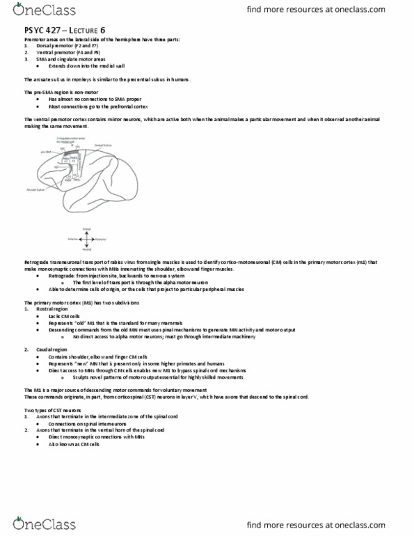
PERIPHERAL MOTOR SYSTEM
Movement is produced by large diameter motor neurons called α MNs that lie in the ventral (anterior) horn of the spinal cord
1. MN axons exit the spinal cord by the ventral root
2. Transverses peripheral nerves
3. Reaches target muscle
The MN branches widely to terminate on 100-1000 individual muscle fibers whose contraction it controls.
• The axon can be seen terminating anywhere between a handful to a large number of muscle fibers in dark red (below)
• The number of terminations determines how finely the α MN can adjust force
Motor unit: ensemble of α MN and the set of muscle fibers it innervates
The muscle fibers are not all together in the same place but are spatially distributed throughout the target muscle. Thus, when the terminating axon discharges, it
produces contraction in multiple parts of the muscle.
Other α MNs innervating the same skeletal muscle are clustered within the same motor nucleus in the ventral spinal cord.
The functional connection between the motor axon and muscle fiber is a chemical synapse called the motor endplate.
An action potential in the neuron results in the release of neurotransmitter acetylcholine, which depolarizes the postsynaptic membrane of the muscle fiber to its
threshold, producing an action potential.
• The inward flow of sodium (Na+) causes depolarization.
• The outward flow of potassium (K+) reverses depolarization.
Muscle fibers are like nerve cells, except unmyelinated. Thus, there is a slowly propagating action potential that spreads in both directions of the muscle fiber.
All muscle fibers innervated by the same MN respond synchronously to each action potential.
• There is simultaneous action in all muscle fibers of a motor unit if threshold is reached
• Recruitment order depends on the size of the α MN, axon diameter, muscle fiber properties, etc.
Above is schematic activity in three different α MN
find more resources at oneclass.com
find more resources at oneclass.com

Motor unit action potential trains (MUAPt): particular pattern or electrophysiological signature of the action potential in muscle fibers
The EMG signal is the aggregation, or addition, of all motor unit action potentials underlying the recording electrodes.
• This makes it possible to back-solve complicated EMG signals to identify individual motor units comprising the total EMG signal
Innervation number: number of muscle fibers per motor unit
• Varies from a minimum of 10 for extraocular muscles (controls eye movement and eyelid elevation) to a maximum of 1000 for lower limb muscles
• Determines how finely force can be graded: the lower the innervation number, the finer the force control
Examples:
Lumbricales or hand muscles involve fine control of movement. Thus, motor units terminate on a small number of muscle fibers in the periphery
• Individual alpha motor neurons produce movement in a small number of muscle fibers, enabling more precise force control
Power muscles, such as biceps, have large innervation numbers because they generate a large amount of force
• The number of alpha motor axons is quite large but the number of muscle fibers is much, much larger
All forms of muscle produce movement by means of active contraction.
Contraction typically results in muscle shortening (concentric contraction), but depending on the action of other muscles and interactions with the environment, one
may observe lengthening during contraction (eccentric contraction).
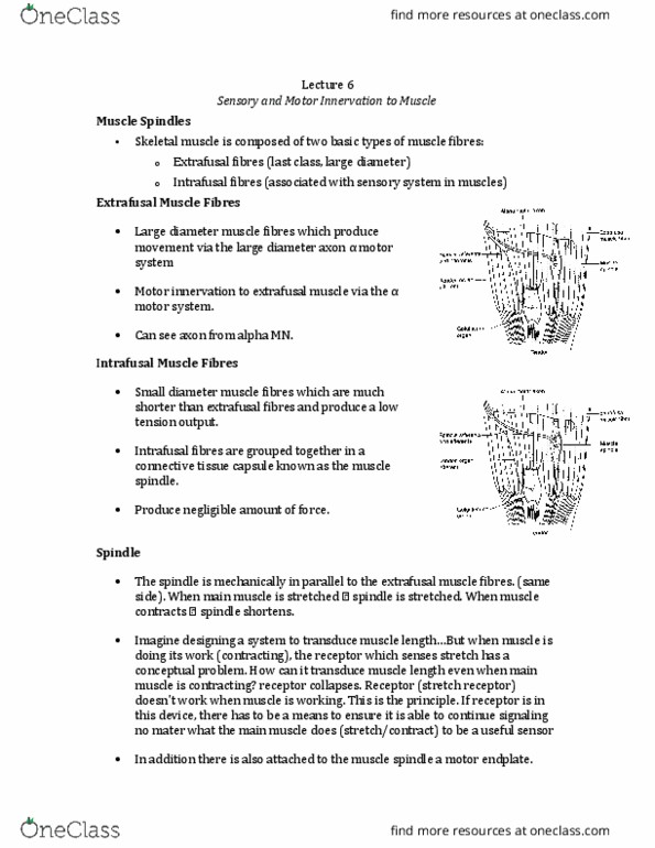
Skeletal muscle (extrafusal muscle): composed of long, thin muscle fibers formed by the fusion of many separate cells
• A contractile machinery: large muscle mass that produces contraction
Three parts of the extrafusal muscle (from smallest to largest:
1. Myofibrils clustered into muscle fibers
2. Muscle fibers clustered into fascicles
3. Fascicles clustered into skeletal muscle
Groups of fibers are bundled into fascicles, which are surrounded by connective tissue
• Muscle fibers are surrounded by fascicles and form compartments within the skeletal muscle
• Myofibrils: the contractile machinery and can be found within individual fibers.
o Contains proteins that produce contraction
Muscle fibers typically terminate in tendinous (collagen) connections to bone with muscle
• Two ends of the muscle that are defined functionally: muscle origin and insertion
find more resources at oneclass.com
find more resources at oneclass.com
Document Summary
Movement is produced by large diameter motor neurons called mns that lie in the ventral (anterior) horn of the spinal cord: mn axons exit the spinal cord by the ventral root. The mn branches widely to terminate on 100-1000 individual muscle fibers whose contraction it controls. The axon can be seen terminating anywhere between a handful to a large number of muscle fibers in dark red (below) The number of terminations determines how finely the mn can adjust force. Motor unit: ensemble of mn and the set of muscle fibers it innervates. The muscle fibers are not all together in the same place but are spatially distributed throughout the target muscle. Thus, when the terminating axon discharges, it produces contraction in multiple parts of the muscle. Other mns innervating the same skeletal muscle are clustered within the same motor nucleus in the ventral spinal cord.