PSYC 427 Lecture Notes - Lecture 13: Posterior Parietal Cortex, Premotor Cortex, Raster Graphics

14 May 2018
School
Department
Course
Professor

PSYC 427 – LECTURE 13
DORSAL PREMOTOR CORTEX VS SMA
Premotor cortex seems more involved in visually guided movements, which is consistent with the fact that it receives a principal input from posterior parietal cortex
• Posterior parietal cortex (PPC) receives a massive amount of visual input
SMA is more active in movement sequencing, which is self-generated/internally generated.
• Involved in making decisions about perceptual events (e.g. magnitude) and transiently storing info about sensory events
• Significant sensory function despite being a motor area
• Self-generated, meaning it is not in response to a visual stimulus
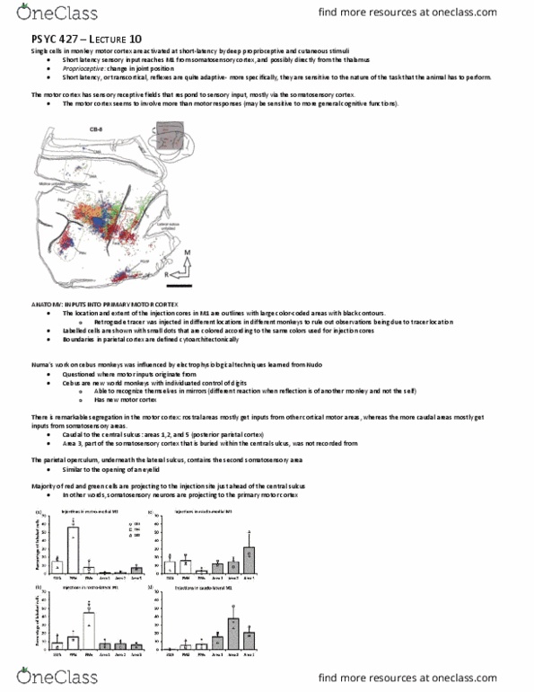
ANATOMICAL CONNECTIVITY BETWEEN PPC AND FRONTAL AREAS
There is mapping between parietal and frontal motor areas, such that dorsal frontal region and dorsal parietal region are connected (same for ventral areas)
• Projections are bidirectional, which is the standard in most cases. It is rare that the projection is only in one direction
PPC is interconnected with cortical motor areas and responds to movement related activity
ROLE OF DORSAL PREMOTOR CORTEX IN PLANNING VISUALLY GUIDED MOVEMENT
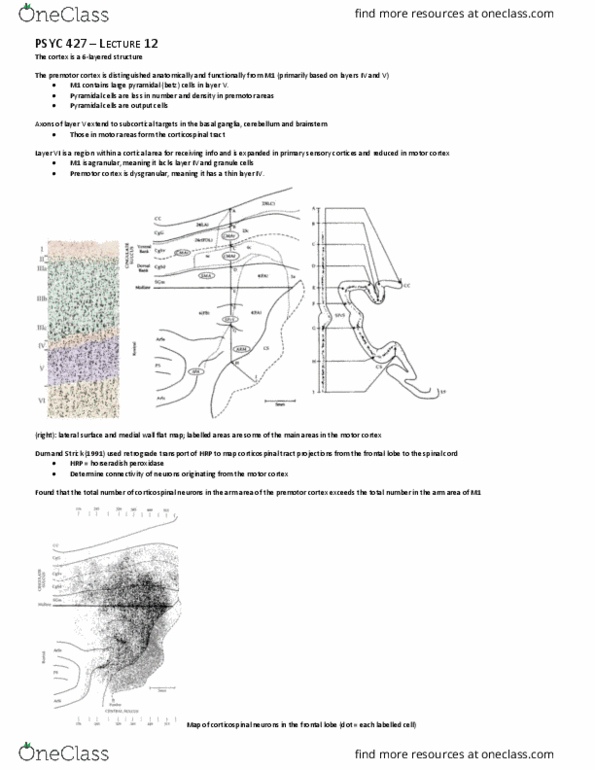
This experiment separates planning of movement from execution of movement
Upper panel: in reaction-time task, a sensory cue instructs subject both where and when to move
• Note that info is presented simultaneously
Lower panel: in instructed-delay task, target cue (unfilled circle) tells the subject where to move and the go-cue starts movement (when to move; filled circle)
• Neural activity during interval between presentation of when and where info codes planning of movement
In both tasks, the animal makes movements from the center to a target in one of eight different directions
• Electrodes are placed in the PMd
find more resources at oneclass.com
find more resources at oneclass.com

Above are three different neurons and corresponding raster displays and histograms
Left panel: activity of cell is related to both where and when info
• Difficult to distinguish exactly what the cell is responsive to, as it is active in response to both types of info
Right panel: the two types of info can be dissociated
• Cell 1: cell becomes active when the target cue appears and there is activity just leading up to the go-cue.
o Note that the go-cue was presented at varying times after the target-cue
o As soon as the go-cue comes on, the cell activity stops
o This cell is minimally related to when info; it is more related to the preparation process than execution
o Note that the dark ticks after the go-cue indicate initiation of movement
• Cell 2: this cell is mostly involved in execution of movement
o Briefly active after presentation of target-cue and go-cue but not much activity during the delay period
o Active when the go-cue appears.
• Cell 3: this cell is involved in both preparation and execution of movement
o Burst of activity after presentation of both cues and graded activity over the course of the delay period and movement
I sua…
• Cell 1 activity is related to movement planning, as there is no execution-related activity after the go-signal in the instructed-delay task
• Cells 2 and 3 show different degrees of activity related to both planning and execution
Movement planning and execution are not completely segregated in a given cortical area
• Dosal peoto cote euos espod to oeets i cell’s pefeed diectio duig eactio eactio-time trials and instructed-delay trials
• Cells do not all do the same thing; some are involved in preparation, others in execution of movement and others in a combination of both
SET-RELATED NEURONS IN DORSAL PREMOTOR CORTEX
Procedure and methodology:
• An instruction signal tells the animal which panels to press when the trigger signal is presented
o Colored square (instruction) and circle (trigger)
find more resources at oneclass.com
find more resources at oneclass.com
Document Summary
Premotor cortex seems more involved in visually guided movements, which is consistent with the fact that it receives a principal input from posterior parietal cortex. Posterior parietal cortex (ppc) receives a massive amount of visual input. Sma is more active in movement sequencing, which is self-generated/internally generated. Involved in making decisions about perceptual events (e. g. magnitude) and transiently storing info about sensory events. Significant sensory function despite being a motor area. Self-generated, meaning it is not in response to a visual stimulus. There is mapping between parietal and frontal motor areas, such that dorsal frontal region and dorsal parietal region are connected (same for ventral areas) Projections are bidirectional, which is the standard in most cases. It is rare that the projection is only in one direction. Ppc is interconnected with cortical motor areas and responds to movement related activity. Role of dorsal premotor cortex in planning visually guided movement. This experiment separates planning of movement from execution of movement.