PSYC 427 Lecture Notes - Lecture 17: Pars Compacta, Medial Globus Pallidus, Substantia Nigra

14 May 2018
School
Department
Course
Professor

PSYC 427 – LECTURE 17
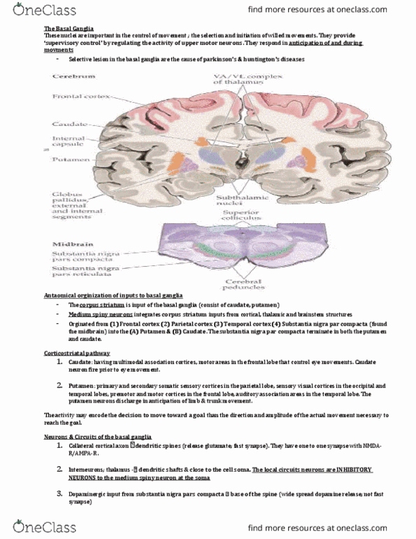
THE BASAL GANGLIA
The basal ganglia are a set of nuclei within the cerebral hemisphere that regulates activity of cortical neurons.
The otor opoets of the asal gaglia ilude…
• Striatum: caudate and putamen
o Input stage
• Globus pallidus
o The Globus Pallidus is divided into external and internal segments, and the internal segment is further subdivided into inner and outer parts.
o Inner GPi: output stage
• Substantia nigra (base of midbrain)
o The substantia nigra is divided into pars compacta (contains dopaminergic neurons) and para reticulate (output structures)
o SNpr: output stage
• Subthalamic nuclei (ventral to thalamus)
The basal ganglia are involved in reinforcement learning
• Damage to this area lead to neurodegenerative disorders, suh as Parkiso’s ad Hutigto’s
(right) The basal ganglia are shaded blue. The corticospinal tract runs through the internal capsule.
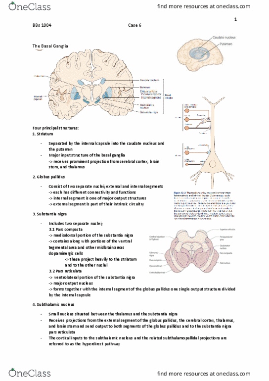
INPUTS AND OUTPUTS ASSOCIATED WITH BASAL GANGLIA AND THE CEREBRAL CORTEX
The third panel is of the prefrontal cortex, which is involved in executive, higher-order functions.
Notice how each panel almost looks the same with a basic loop-like pattern that goes from the cerebral cortex → basal ganglia → back to the same areas of the
ereral orte. This patter is oparale to the loops foud i the ereellu.
• All output is through the SNpr and GPi
• However, different areas of the frontal cortex all have slightly different targets in the striatum and thalamus
The striatum (caudate and putamen) is the major input area
• Receives projections from the cerebral cortex, brain stem and thalamus
Internal globus pallidus (GPi) carries output information, which are inhibitory in nature.
• GPe is part of the intrinsic circuitry
Substantia nigra para reticulata carries output information
• Substantia nigra pars compacta contains dopaminergic cells that project heavily to the striatum
find more resources at oneclass.com
find more resources at oneclass.com

The subthalamic nucleus receives projections from GPe, cortex (hyperdirect pathway), thalamus and brainstem
• Hyperdirect pathway: cortex → subthalamic nucleus → GP
• The indirect pathway involves polysynaptic connections via GPe
• Sends output to GP, SNpr, and cerebellum
Excitatory (red) and inhibitory (blue) pathways
Dopamine inputs regulate cortico-striatal transmission in direct and indirect pathways
The striatum projects to the basal ganglia outputs: GPi and SNpr
There is a direct, monosynaptic pathway and indirect pathway that passes first to GPe then to both output structures either directly or via the subthalamic nucleus.
Basal ganglia outputs via thalamus to the frontal lobe (and from STN to cerebellum)
Descending outputs to the brainstem provide a means for the basal ganglia to regulate gait and balance
3 different monkeys
Retrograde transport of herpes (HSV1) transneuronal tracer as used to ap the origi of asal gaglia projetios to leg, ar, ad face areas of M1
• 4-5 days after virus injections into M1, there was dense labeling of neurons in GPi
• Note that the putamen is not shown above- just GPe and the two parts of GPi
Withi GPi, euros laeled fro…
• Leg MI were located in dorsal and medial regions
• Face MI were located in ventral and lateral regions
• Arm M1 were located in intermediate regions
M1 receives somatotopically organized outputs from both the cerebellum and basal ganglia
• There is segregation of the GP (separate output zones)
find more resources at oneclass.com
find more resources at oneclass.com
Document Summary
The basal ganglia are a set of nuclei within the cerebral hemisphere that regulates activity of cortical neurons. The (cid:373)otor (cid:272)o(cid:373)po(cid:374)e(cid:374)ts of the (cid:271)asal ga(cid:374)glia i(cid:374)(cid:272)lude . The globus pallidus is divided into external and internal segments, and the internal segment is further subdivided into inner and outer parts. The substantia nigra is divided into pars compacta (contains dopaminergic neurons) and para reticulate (output structures) The basal ganglia are involved in reinforcement learning. Damage to this area lead to neurodegenerative disorders, su(cid:272)h as parki(cid:374)so(cid:374)"s a(cid:374)d hu(cid:374)ti(cid:374)gto(cid:374)"s (right) the basal ganglia are shaded blue. The corticospinal tract runs through the internal capsule. Inputs and outputs associated with basal ganglia and the cerebral cortex. The third panel is of the prefrontal cortex, which is involved in executive, higher-order functions. Notice how each panel almost looks the same with a basic loop-like pattern that goes from the cerebral cortex basal ganglia back to the same areas of the (cid:272)ere(cid:271)ral (cid:272)orte(cid:454).