HTHSCI 1DT3 Lecture 4: 1.1 Cells.10

23 Jun 2018
School
Department
Course
Professor

Development: Anita Hall’s lectures
Growth/renewal: Lectures on Regeneration; Stem Cells
Morphology & Intracellular architecture: Introductory sections of most neuro text
books; Alberts.
Neuronal Polarity & Trafficking. (2003). Horton & Ehlers. Neuron 40, 277-295.
Astrocytes and Radial Glia
Glia can be subdivided into:
Macroglia – (Schwann cells, oligodendrocytes, astrocytes/radial glia, olfactory
ensheathing cells)
Microglia
Development of Astrocytes
CNS glia arise from the ventricular zone of the neural tube (similar to CNS neurons). C.f.
Schwann cells that arise from the neural crest (as do PNS neurons)
Astrocytes and neurons arise from common progenitor cells in the neuroepithelium (perhaps
even radial glia?)
Astrocyte Cell Biology
Astrocytes can be:
FiBROUS (TYPE II)
These contain much fibrous material (GFAP) and are prevalent in myelinated nerve
bundles in the white matter. These astrocytes are star shaped.
PROTOPLASMIC (TYPE I)
These have less GFAP and are abundant in the grey matter around cell bodies, synapses
and dendrites.
GFAP – an intermediate filament protein that is expressed in astrocytes and radial glia. It is a useful
marker for these cells.
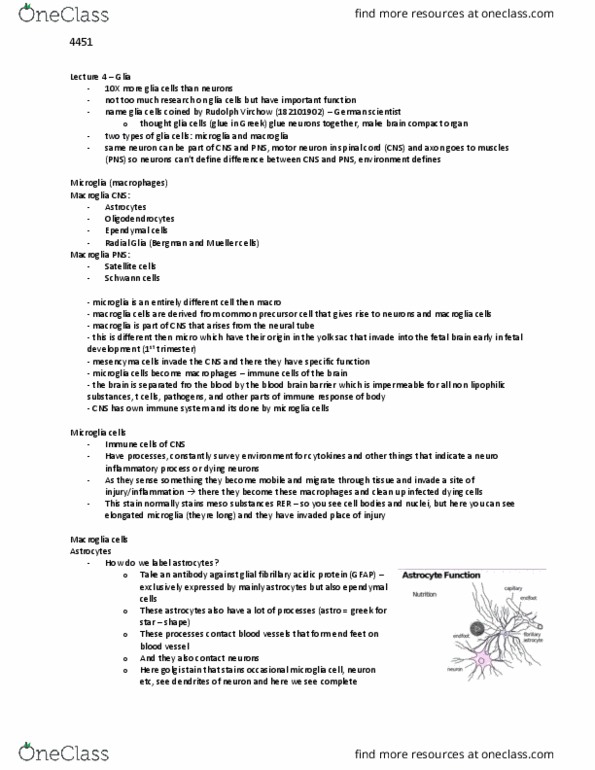
In the brain and spinal cord, astrocytes have:
Many thin processes around:
Capillaries (top left on diagram)
Synapses
Surfaces of neurons
Cytoskeleton
Intermediate filament (GFAP)
Microtubules
Actin
Type II Astrocytes are
more fibrous.
Glycogen granules (large energy
stores)
Rough ER, Golgi Apparatus
Large Nucleus
Watery Cytoplasm
Functions of astrocytes and radial glia
Astrocytes interact intimately with neurons
o
o
o
•
•
o
o
•
•
•
o
o
•
o
•
•
•
o
•
•
•
•
o
o
o
o
•

Astrocytes form large syncytial interconnected networks coupled by gap junctions
This has been shown by injecting dyes into a single astrocyte and seeing the dye spread through
the network.
Gap junctions allow exchange of ions and small molecules <1kDa.
Functions of Astrocytes:
At blood brain barrier, they act as a gateway between general circulation and the neurons of
the CNS (neurovascular unit)
The blood-brain barrier is formed by very tight associations of brain capillary
endothelial cells.
It is essential for controlling entry of molecules and ions from the general circulation
into the central nervous system.
Astrocytic endfeed wrap endothelial cells, providing a gateway for nutrients into the
CNS and removal of metabolites from the CNS.
•
•
•
1)
o
o
o

Provide Energy – to neurons in the form of lactate produced from glycogen specifically in
astrocytes.
Astrocytes are the only cells in the brain that store glycogen (hence the large glycogen
granules in the cytoplasm) – this is enough to last for tens of minutes.
Removal of Potassium Ions – from extracellular space. Essential for neuronal function, since
extracellular potassium concentration must be kept low.
After intense neuronal activity, local concentrations of potassium can become very high.
Potassium is redistributed from these regions to other regions, by transport through the
astrocytic network via gap junctions.
Astrocytes therefore act as a potassium reservoir, maintaining a good supply of
potassium but keeping it away from the extracellular space until required.
Provide Structural Support
Sturdy star shape
Lots of GFAP (high tensile strength)
Guidance for neuronal development (radial glia)
Radial glia span the cortex radially from the inner to outer layers
They are important for neuronal migration.
Examples include Bergmann glia in the cerebellum and the Muller cells in the retina
Note the similarity to neurepithelial cells during neurogenesis.
Cortical neurons migrate up the radial glia processes during cortical development.
They crawl over the glial cell surface using homophilic binding cell adhesion molecules
of the immunoglobulin superfamily. (Anita Hall’s Lecture / Molecules & Mechanisms
of Regeneration)
As well as directing migration of neuron resulting from neurogenesis, glial cells also
provide guidance for migrating growth cones during axonal outgrowth towards the
target cell.
The same molecules and mechanisms underlie both migrations.
Guidance from glial cells provides the pathway guidance using pathway guidance cues
(on cell/matrix) – helps guides migration of axonal growth cones to target.
Soluble Chemoattractants
Soluble survival factors
Provides TROPHIC (Survival) Factors to neurons !
(e.g. glial cell derived growth factor – GDNF) / Promising clinical trials for Parkinson’s
disease.
Water Reservoir in the CNS – As astrocytes have a lot of watery cytoplasm with relatively few
organelles, they can act as a water reservoir in the CNS.
Act as PHAGOCYTES to REMOVE neuronal debris from within the CNS and transport it
back to the bloodstream.
Regulate CONCENTRATION of NEUROTRANSMITTER
e.g. take up glutamate or GABA, convert to glutamine and release to be taken up by neurons
(that convert it back to glutamate to be reused)
2)
o
3)
o
o
o
4)
o
o
5)
o
o
o
o
o
o
o
o
o
•
•
6)
o
7)
8)
9)
Document Summary
Morphology & intracellular architecture: introductory sections of most neuro text books; alberts. Macroglia (schwann cells, oligodendrocytes, astrocytes/radial glia, olfactory ensheathing cells) Cns glia arise from the ventricular zone of the neural tube (similar to cns neurons). Schwann cells that arise from the neural crest (as do pns neurons) Astrocytes and neurons arise from common progenitor cells in the neuroepithelium (perhaps even radial glia?) These contain much fibrous material (gfap) and are prevalent in myelinated nerve bundles in the white matter. These have less gfap and are abundant in the grey matter around cell bodies, synapses and dendrites. Gfap an intermediate filament protein that is expressed in astrocytes and radial glia. It is a useful marker for these cells. In the brain and spinal cord, astrocytes have: o. Type ii astrocytes are more fibrous. o o o o. Astrocytes form large syncytial interconnected networks coupled by gap junctions.