HTHSCI 2FF3 Lecture Notes - Lecture 2: Indometacin, Ductus Venosus, Brachiocephalic Artery

1 Nov 2016
School
Department
Course
Professor
Document Summary
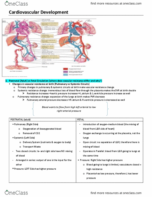
In a fetus, the placenta is the gas exchange organ (instead of the lungs). It is supplied by branches of the aorta. Thus, de-oxygynated blood needs to travel from the right side to the left side of the heart, to get to the placenta through the aorta. Foramen ovale: a hole in the atrial septum: ductus arteriosus: a tube connecting the. Note, these structures are not present in an adult heart. Blood flow follows the path of least resistance. In fetal state, pulmonary arteries are constricted, thus resistance and pressure are consequently high in the right side. In contrast, the left side of the heart pumps to the systemic circulation of low resistance. Consequently, pressure in pulmonary arteries is higher than in aorta. Thus, blood is pushed right to left through the formaen ovale, and the ductus arteriosus into the aorta. Because the lungs are bypassed, this right to left redirection of blood flow is called a shunt .