KINESIOL 1A03 Lecture Notes - Lecture 15: Pseudostratified Columnar Epithelium, Vital Capacity, Cough Reflex

15 Apr 2018
School
Department
Course
Professor
Document Summary
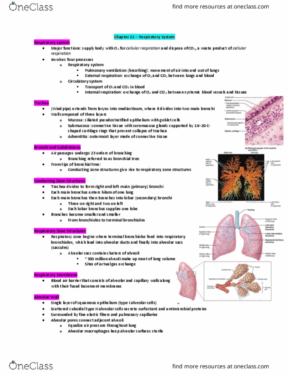
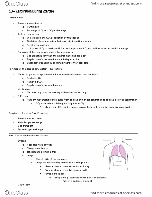
Functional anatomy respiratory zone: site of gas exchange: microscopic structures: respiratory bronchioles, alveolar ducts and alveoli conducting zone: conduits to gas exchange sites includes all other respiratory structures respiratory muscles: diaphragm and other muscles that promote ventilation. Bronchi and subdivisions air passages undergo 23 orders of branching branching pattern called the bronchial (respiratory) tree. Alveoli respiratory bronchioles, alveolar ducts, alveolar sacs (clusters of alveoli) Pleurae thin, double-layered serosa parietal pleura on thoracic wall and superior face of diaphragm visceral pleura on external lung surface pleural fluid fills the slit-like pleural cavity: provides lubrication and surface tension. Ppul rises (to +1 mmhg) air flows out of the lungs down its pressure gradient until ppul = 0: **note: forced expiration is an active process: it uses abdominal and internal intercostal muscles. Intrapulmonary pressure intrapulmonary (intra-alveolar) pressure (ppul: pressure in the alveoli fluctuates with breathing, always eventually equalized with patm.