MEDRADSC 2I03 Lecture 15: Pathologies of the Lower Respiratory Tract

11 Dec 2018
School
Department
Course
Professor
Document Summary
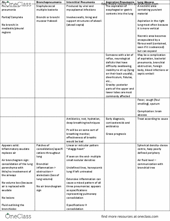
Sunday, november 18, 2018 9:33 pm: lower respiratory, diseases of lung! Pneumonia: acute pneumonia - inflammation of lung, three basic radiographic patterns -> three different areas, alveolar pneumonia, bronchopneumonia. Alveolar pneumonia - air broncho sign: alveolar pneumonia. Inflammatory exudate replaces air: components produced from infection. Can involve pulmonary segments or entire lobe: depends on disease process b/w pts. No bronchi in mediastinal or pleural regions: rule out lesions. Portion of right bronchus splitting: classic sign of alveolar pneumonia. Air broncho sign: everything but the bronchus filled w fluid, bronchus (air) contrast w lung, all looks v hazy, can see where left main bronchus comes into lung. Normal lung: portion of right bronchus splitting (black arrows, consolidations on mid-right lung, normal lung laterally, only portions of right lung affected, left lung unaffected. Bronchopneumonia - ill defined consolidation at right base, scattering: bronchopneumonia. Bronchial inflammation -> obstructs airway -> atelectasis. Volume loss: bc of atelectasis, but also increased density.