MEDRADSC 2Z03 Lecture Notes - Lecture 11: Soft Tissue, Talar, Hyaline Cartilage

29 Jun 2016
School
Department
Course
Professor
Document Summary
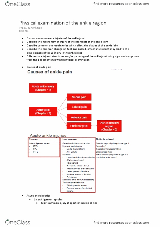
Common indications for mri of the foot and ankle include: Figure 1 below indicates the major anatomical structures and tissues within the foot, ankle and lower leg. Figure 2, below, indicates the planes used for transverse and coronal images of the ankle and foot. Structures of the foot that are commonly examined by mri include: In figure 3, below, are three images of a patient who sustained an inversion injury (sprain) of the ankle. The sagittal and axial images demonstrate the high signal of a moderately sized joint effusion (swelling of the joint). In the coronal image, some high signal can be seen in the talar dome. This is a small bone bruise caused by bone hitting against bone during the twisting motion. The images in figure 4, below, show a normal achilles tendon in a t1-weighted image and t1 and t2-weighted images of a subject with a full thickness rupture of the tendon.