MEDRADSC 3I03 Lecture Notes - Lecture 15: Inferior Colliculus, Cerebellar Peduncle, Middle Cerebellar Peduncle

7 Jan 2017
School
Department
Course
Professor
Document Summary
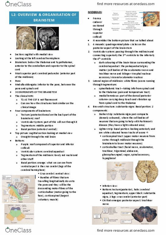
Tectmentum holds in the reticular formation (not seen: nuclei cell bodies. Tectum at the same spot, posterior part of the midbrain: aka quadrigeminal plate. Little dot through the tectum, cerebral aqueduct. Thalamus builds the side wall of the ventricle. Dark space posterior to tectum = quadrigeminal cistern. Interpedunclar cistern = between the two peduncles. Anterior to the pons, tiny white dot = basilar artery. The first label we see is the middle cerebral peduncle. Middle cerebellar peduncle attaches cerebellum to pons. Inferior cerebellar peduncle to medulla oblongata. The ventral portion of the pons is labelled as basilar portion of pons. Creating the inferior portion of the clivius. Optic foramina in the lesser wing of sphenoid. Middle of it would be the thickest, White is myelin sheath tiny bones in the ear, two nerves are in there, Vii is sup and viii is the inf one. Cp angle pathology going in the semicircular canal of the ear bones.