NURSING 1J02 Lecture Notes - Lecture 7: Common Iliac Artery, Colic Flexures, Rectus Abdominis Muscle

2 Mar 2017
School
Department
Course
Professor

Document Summary
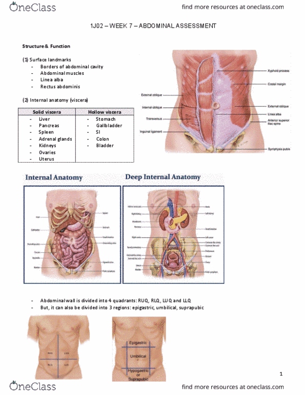
Abdomen: large oval cavity from diaphragm to top of pelvis. Bordered in the back by vertebral column & paravertebral muscles & at sides & front by lower rib cage. 4 layers of large, flat muscles form ventral abdominal wall, joined together by the linea alba. Muscles protect & hold organs in place and flex vertebral column. All organs in abdominal cavity are called viscera. Solid viscera: maintain a characteristic shape (kidney, pancreas, spleen, adrenal glands, liver, ovaries & uterus) Liver fills up most of ruq & extends over left midclavicular line. Lower edge of liver & right kidney may normally be palpable. Ovaries palpable only in bimanual assessment during pelvic examination. Hollow viscera: shape depends on contents (stomach, gallbladder, small intestine, colon & bladder) - Stomach just below diaphragm (b/w liver & spleen) Spleen- soft mass of lymphatic tissue on posterolateral wall of abdominal cavity behind & parallel to.