ANAT 216 Lecture Notes - Lecture 11: Superior Mesenteric Vein, Superior Mesenteric Artery, Inferior Mesenteric Artery

28 Apr 2018
School
Department
Course
Professor

ANAT 216 week 4 lecture 3
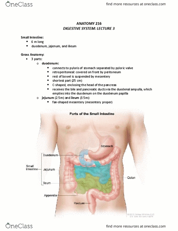
Small
intestine
6-7m for digestion and absorption (90%),
wrapped in mesentery proper (peritoneum)
except duodenum from posterior ab wall
Microvilli move by supporting
microfilaments, villi move by muscularis
mucosa, pilcae drainage move by muscularis
mucosa and externa
Duodenum: 25cm, C-shaped to enclose head
of pancreas, retroperitoneal, receives bile,
chyme via duodenal ampulla
Bruer’s glads: serete alkalie uus to
neut chyme
Jejum: 2.5m fan shaped mesentery
Ileum: opens into cecum at ileocecal valve
which prevent backflow into SI
Peyer’s pathes: aggregatios of lyph
tissues
Small
histology
Mucosa: vili covered by simple columnar
(enterocytes) which are covered by microvilli
(brush border) and Goblet cells supported by
lamina propria (dense irreg w cap networks
and lymph lacteals, forms core of villus,
otais Peyer’s pathes)
Intestinal glands: crypts of Lieberkuhn spaces
b/n packed tubular folds, entrance for bush
border enzymes, opening from intestinal
glands to lumen at base of vili
Absorptive, goblet (lub chyme), Paneth
(defensive, prod lysozymes), enteroendocrine
(scattered, prod CCK, gastrin, VIP, secretin),
stem cells
Suuosa: Bruer’s glads i duodeu
All parts have plicae circulates/circular folds=
permanent folds
Long folds: disappear as digestive tract fills,
allow expansion
Blood
supply of
small
Superior mesenteric artery from ab aorta
Superior mesenteric vein which drains into
hepatic portal vein
Large
intestine
1.5m, horseshoe-shaped, lies distal to
stomach and liver
Cecum: ileocecal valve (attachment to ileum)
Vermiform appendix: slender hollow passage
(9cm) filled w lymphoid nodule and attached
to cecum
Colon: largest part
find more resources at oneclass.com
find more resources at oneclass.com
Document Summary
6-7m for digestion and absorption (90%), wrapped in mesentery proper (peritoneum) except duodenum from posterior ab wall. Microvilli move by supporting microfilaments, villi move by muscularis mucosa, pilcae drainage move by muscularis mucosa and externa. Duodenum: 25cm, c-shaped to enclose head of pancreas, retroperitoneal, receives bile, chyme via duodenal ampulla. Bru(cid:374)(cid:374)er"s gla(cid:374)ds: se(cid:272)rete alkali(cid:374)e (cid:373)u(cid:272)us to neut chyme. Ileum: opens into cecum at ileocecal valve which prevent backflow into si. Mucosa: vili covered by simple columnar (enterocytes) which are covered by microvilli (brush border) and goblet cells supported by lamina propria (dense irreg w cap networks and lymph lacteals, forms core of villus, (cid:272)o(cid:374)tai(cid:374)s peyer"s pat(cid:272)hes) Intestinal glands: crypts of lieberkuhn spaces b/n packed tubular folds, entrance for bush border enzymes, opening from intestinal glands to lumen at base of vili. Absorptive, goblet (lub chyme), paneth (defensive, prod lysozymes), enteroendocrine (scattered, prod cck, gastrin, vip, secretin), stem cells. All parts have plicae circulates/circular folds= permanent folds.