ANAT 216 Lecture Notes - Lecture 12: Portal Vein, Common Bile Duct, Hepatic Veins

28 Apr 2018
School
Department
Course
Professor

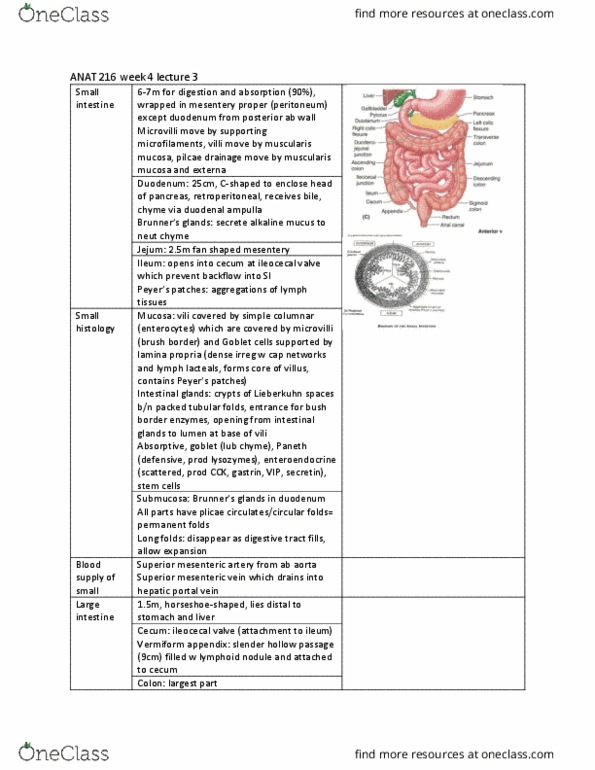
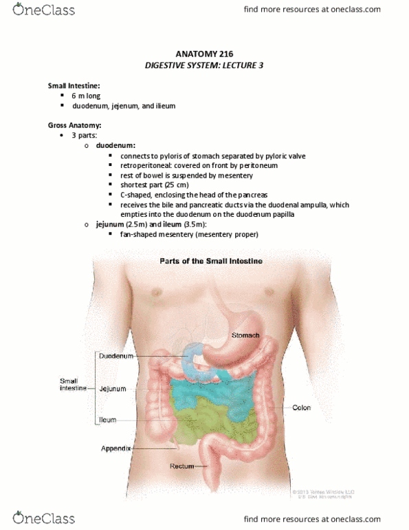
ANAT 216 week 5 lecture 1
Large
histology
Muscoa: no plicae circularis or villi bc little
absorption here
Simple columnar w same cells as small but
Goblet cells increase for lub
Strat squamous in anal canal, deeper
intestinal glands (crypts), accumulations of
lymphatic tissue in lamina propria and
submucosa
Submucosa: normal
Muscularis externa:
Taeniae: outer long layer made of 3
smooth bands
Hasutra: series of pouches in wall of colon,
created by muscle tone in taeniae, permit
elongation and expansion
Circular muscle thickens to form sphincter
in anal canal
Liver
Starts at 5th rib9th largest visceral organ
(1.5kg), wrapped in fibrous capsule
Diaphragmatic (superior & anterior) and
visceral (posterior) surfaces
R largest lobe, R+ L lobe on superior
surface, caudate located left to fossa of
vena cava on posterior surface, quadrate
located next to gall bladder fossa on
inferior surface below caudate
Peritoneum: completely covered except
base area b/n 2 coronary lig, visceral-
stomach, duodenum, gall bladder, R kidney
+ R colic flexure
Falciform lig: peritoneum, attaches liver to
anterior ab wall, sep R and L lobes and
make coronary lig, reminant of umbilical
vein
Round lig: fibrous band in free margin of
falciform lig
Coronary lig: contains bare area, suspend
liver from inferior diaphragm surface
Lesser omentum
find more resources at oneclass.com
find more resources at oneclass.com
Document Summary
Muscoa: no plicae circularis or villi bc little absorption here. Simple columnar w same cells as small but. Strat squamous in anal canal, deeper intestinal glands (crypts), accumulations of lymphatic tissue in lamina propria and submucosa. Taeniae: outer long layer made of 3 smooth bands. Hasutra: series of pouches in wall of colon, created by muscle tone in taeniae, permit elongation and expansion. Circular muscle thickens to form sphincter in anal canal. Starts at 5th rib 9th largest visceral organ (1. 5kg), wrapped in fibrous capsule. Diaphragmatic (superior & anterior) and visceral (posterior) surfaces. R largest lobe, r+ l lobe on superior surface, caudate located left to fossa of vena cava on posterior surface, quadrate located next to gall bladder fossa on inferior surface below caudate. Peritoneum: completely covered except base area b/n 2 coronary lig, visceral- stomach, duodenum, gall bladder, r kidney.