MICR 221 Lecture Notes - Lecture 11: Osmotic Shock, Crystal Violet, Thermoacidophile

12 Nov 2016
School
Department
Course
Professor
Document Summary
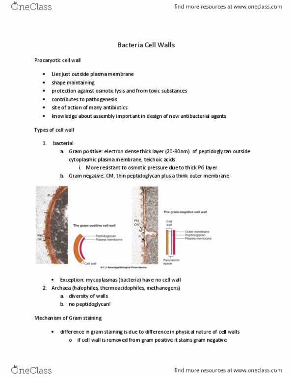
Site of action of many antibiotics knowledge of assembly important in design of new antibacterial agents. Prokaryotic cell wall types: bacterial, gram positive cm +thick electron layer. Peptidoglycan to techoic acids: gram negative cm + thin peptidoglycan + om (lps, proteins, phospholipids). Bacteriophage use lps as a receptor to bind the cell: mycoplasmas (cid:374)o (cid:272)ell (cid:449)all. Ie) pe(cid:374)i(cid:272)illi(cid:374) (cid:449)ill ha(cid:448)e (cid:374)o effe(cid:272)t o(cid:374) these. No wall therefore sensitive to osmotic pressure (protected by sterols in cm). Associated with bacteria causing pneumonia: archea (halophiles, thermoacidophiles, methanogens) Gram positives have more peptidoglycan which takes up the crystal violet stain better than gram positives. Also, gram positives have lipids and proteins in their outer layer which prevents uptake of the stain. Barrier to keep periplasmic proteins from diffusing away. Relatively permeable to small hydrophilic molecules (ie sugars) Found in outer leaflet of lipid bilayer of om. Ie) endotoxin caused fever, diarrhea, interferes with blood clotting.