BIOCH200 Lecture Notes - Lecture 8: Peptide, Chief Operating Officer, Coordinate Covalent Bond

5 May 2018
School
Department
Course
Professor

Introductory Biochemistry; Hemoglobin and Myoglobin
Function
❏Determined absolutely by its structure → Primary Structure.
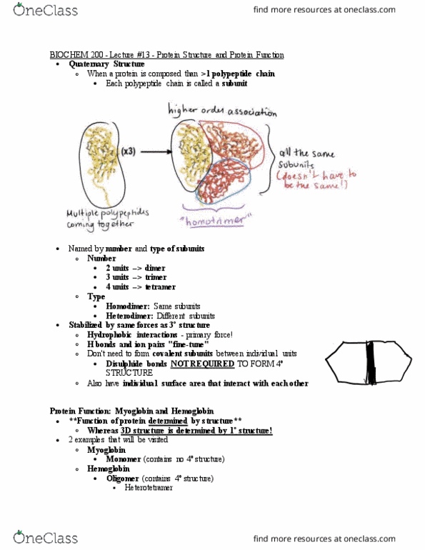
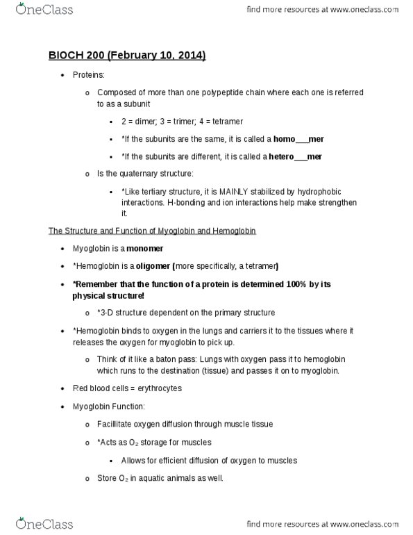
Myoglobin
❏Monomer → 1 subunit (polypeptide) and 1 domain
❏Binds oxygen in the muscle
❏Hydrophobic pocket between helix E and F
❏8 Alpha helices and irregular structures w/ a heme prosthetic group.
❏Helix A = N Terminus
❏Helix H = C terminus
❏Does not have Quaternary Structure
❏Binds 1 Oxygen
Hemoglobin
❏Oligomer → more than 1 subunit and each subunit has one domain
❏2 Alpha subunits and 2 beta subunits
❏Each subunit has 8 alpha helices, loops and 1 heme.
❏Binds to oxygen in the lungs and releases it in the tissues
❏Has Quaternary Structure
❏Binds 4 Oxygen
Function of Myoglobin
❏Stores oxygen in aquatic animals
❏Act as local reserve of oxygen during intense exercise
❏Facilitate oxygen diffusion through muscle tissues.
Function of Proteins
❏Dependent on ability to bind other small molecules (ligands) reversibly.
❏The greater the affinity of Protein X for Y the more XY that is produced.
❏If affinity is low then Kd is high.
❏If affinity is high then Kd is low (substrate binds to protein).
Ligand Binding Curves
❏Thick arrow → High affinity
❏Thin arrow → Low affinity
Structure of Heme
❏Circular, planar and heterocyclic → Porphyrin ring contains Fe2+ ion coordinated (one atom
donates both electrons to atom) between 4 Nitrogens.
❏Contains two propionyl groups (COO-) that are polar (remaining is non-polar) and allow heme to
orient itself.
❏6th coordination bond → Distal Histidine (E7) assists oxygen binding through hydrogen bonding
with oxygen. → Further away than F8.
❏5th coordination bond → Proximal Histidine (F8) → Permanent bond, and close to Fe2+
❏Porphyrin ring (Heme) held by hydrophobic interactions and coordination bond between HisF8
and Fe2+
Cofactors
❏Non-protein chemical compound required for proteins biological activities.
❏Example: Heme.
Binding Sites
❏Optimize specificity and affinity.
Document Summary
Determined absolutely by its structure primary structure. Monomer 1 subunit (polypeptide) and 1 domain. Hydrophobic pocket between helix e and f. 8 alpha helices and irregular structures w/ a heme prosthetic group. Oligomer more than 1 subunit and each subunit has one domain. 2 alpha subunits and 2 beta subunits. Each subunit has 8 alpha helices, loops and 1 heme. Binds to oxygen in the lungs and releases it in the tissues. Act as local reserve of oxygen during intense exercise. Dependent on ability to bind other small molecules (ligands) reversibly. The greater the affinity of protein x for y the more xy that is produced. If affinity is low then kd is high. If affinity is high then kd is low (substrate binds to protein). Circular, planar and heterocyclic porphyrin ring contains fe2+ ion coordinated (one atom donates both electrons to atom) between 4 nitrogens.