ANSC 3080 Lecture Notes - Lecture 20: Cardiac Cycle, Systolic Geometry, Chordae Tendineae

27 Mar 2018
School
Department
Course
Professor
Document Summary
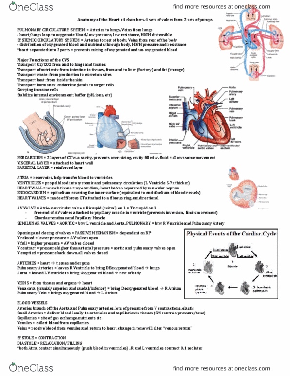
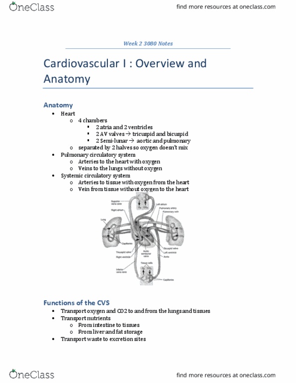
2 layers of connective tissue with a cavity. Cavity filled with fluid to allow movement. 2 atria that collect blood from venous return. 2 ventricles the pump blood into systemic and pulmonary circulation. Septum separates the heart into 2 halves. Valves made of fibrous ct attached to a fibrous ring. Opening and closing of valves are passive, based on blood pressure. Tricuspid (right), bicuspid/mitral (left) are stabilized by chordae tendinae connected to papillary muscle in the ventricles to prevent back flow. Aortic and pulmonary semilunar valves are stronger so they do not need chordae tendinae. Transport gases, nutrients (from intestine, liver, fat), waste, heart, hormones. Carry immune cells and drop them off at the infection site especially neutrophil. Stabilize internal environment as buffer to control ion concentration. Branch off to arterioles and then capillary beds. Contraction of smooth muscle can control blood pressure. Superior and inferior vena cava bring blood to the right atrium.