FRHD 2100 Lecture Notes - Lecture 8: Brush Border, Lipoprotein, Glycogen

1 Jun 2018
School
Department
Course
Professor

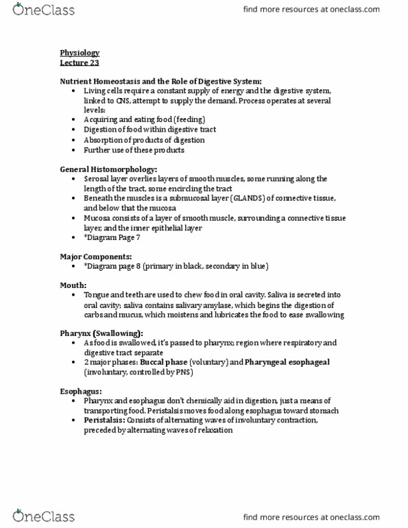
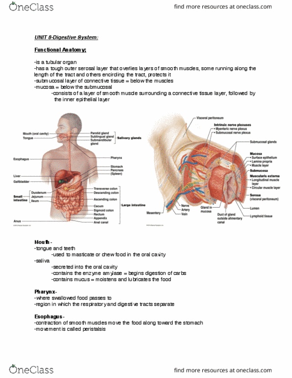
FUCNTIONAL ANATOMY OF THE DIGESTIVE TRACT
• Mouth to anus
• Tubular organ
• Tough outer serosal layer
o Overlies layers of smooth muscle running along length, encircling the tract
The Mouth
• Starts in the mouth with tongue and teeth
o Used to chew food
• Saliva
o Contains amylase (enzyme)
o Begins digestion of carbs and mucus
o Moistens and lubricates food
• As food is swallowed, it passes to the pharynx
o Where respiratory and digestive tracts separate
The Esophagus
• Form pharynx into esophagus
• Contraction of smooth muscle move food toward stomach
o Peristalsis
The Stomach
• Cardiac region
o Receives food from esophagus
• Pyloric region
o Delivers partially digested product to next section of digestive tract
• Duodenum
o First section of small intestine
• Gastric glands in stomach secrete acidic gastric juice
• Acid prevented from reaching esophagus by anterior sphincter
• Chyme
o Acidic stomach content
o Released in small spurts into duodenum by opening second sphincter
▪ Posterior
• Same smooth muscle layers found in stomach
• Smooth muscle found in wall of stomach running
o Length of the organ
o Around the organ
o Run in oblique direction
o Muscle act to mix and mechanically break up food
• Gastric glands
o Embedded in pits
o Cells that produce secretions that assist with digestion
find more resources at oneclass.com
find more resources at oneclass.com

• Parietal cells
o Secrete hydrochloric acid
▪ Very acidic – pH 2
• Chief cells
o Secrete pepsin that digests protein
• Mucous secreting cells
o Secrete sticky alkaline mucus
o Protects stomach lining from acid burn
• Carb digestion
o Continues in anterior stomach
• Protein digestion
o Begins in posterior region
• Small molecules (water, alcohol, aspirin) absorbed in stomach
The Small Intestine
• Chyme moves from stomach into duodenum
o By opening posterior sphincter
o Immediately neutralized by alkaline bile
• Duodenum – 5%
• Jejunum – 40%
• Ilium – 55%
• Alkaline bile
o Released from gall bladder
o Brought to duodenum via bile duct
o Bile is synthesized by liver and stored in gall bladder
o Bile neutralizes chyme and makes fat digestion more efficient
• Pancreatic juices
o Released into duodenum
o Juices are mix of enzymes that digest carbs, small proteins, and lipids
• Large surface area of mucosa makes digestion and absorption possible
• Structural modifications of small intestine
o Folding of mucosa
▪ Structures known as ridges
▪ Made up of finger-like villi
▪ Surface of villi or absorptive cells are microvilli
• Aka brush border
o Lacteals essential in absorption of lipids
o Presence of villi and microvilli
• Secretions DO NOT contain enzymes
• Digestive enzymes are located on brush border
• End products
o Amino acids (protein)
o Glycerol and fatty acids (lipids)
find more resources at oneclass.com
find more resources at oneclass.com
Document Summary
Fucntional anatomy of the digestive tract: mouth to anus, tubular organ, tough outer serosal layer, overlies layers of smooth muscle running along length, encircling the tract. The esophagus: form pharynx into esophagus, contraction of smooth muscle move food toward stomach, peristalsis. The stomach: cardiac region, receives food from esophagus. The small intestine: chyme moves from stomach into duodenum, by opening posterior sphincter. Immediately neutralized by alkaline bile: duodenum 5% Juices are mix of enzymes that digest carbs, small proteins, and lipids. The large intestine: from ilium to colon/large intestine, produces a lot of mucus, lubricates, acts as colony of bacteria, valve prevents contents from colon from back flowing. Lipid digestion: triglycerides, phospholipids, sterols/cholesterol, not water soluble, enzymes have limited access to molecules of insoluble lipid, bile salts help lipid digestion. Lipids can be broken down into small droplets. Lipase: present in pancreatic juices, occurs in small intestine.