ZOO 3200 Lecture Notes - Lecture 5: Faraday Constant, Electromotive Force, Axon Hillock

2 May 2018
School
Department
Course
Professor

Structure-Function Relationship of Neurons
!
Glial Cells
!
Electrical Signals in Neurons
!
The Nernst Equation
!
The Goldman Equation
!
Electrochemical potentials
○
Membrane Potential -mathematical underpinnings
!
Outline:
Readings: Chapter 12 (pages 295-308), Invertebrate Studies and their
Contribution to Neuroscience
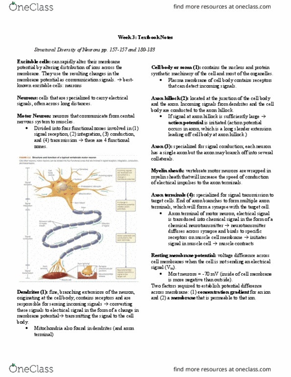
Neuron -anatomical and functional unit of the nervous system
Signal reception (input): dendrites
○
Signal integration: axon hillock
○
Signal conduction: axon
○
Signal transmission (output): axon terminals
○
Four functional zones:
!
Synapse: connection between two nerves or a nerve cell and
muscle cell
!
Cell body (soma) to axon terminals
○
Neural signals only travel in one direction
!
MS patients lose their myelin sheaths which causes signal
strength to decrease and other complications
○
Myelin sheath insulates the neuron
!
Structure-Function Relationship of Neurons
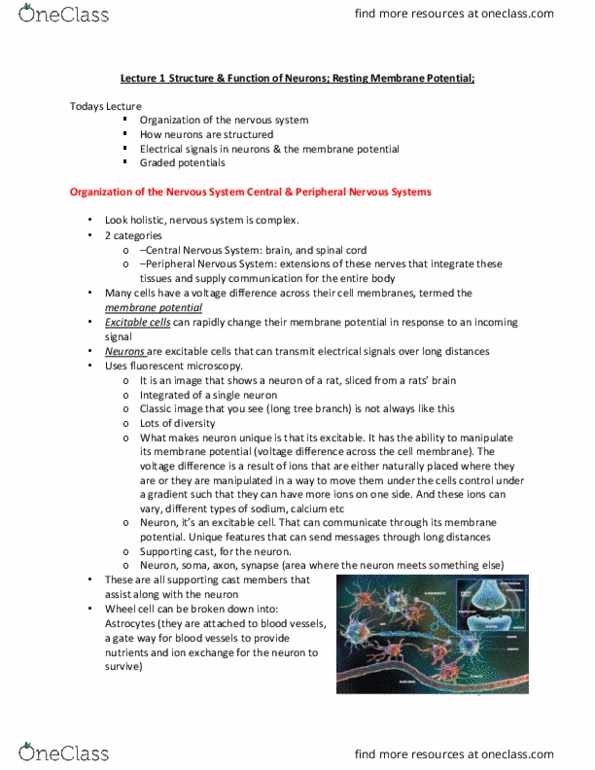
Most neural cells are glial cells
!
Glial cells support neurons
!
They do not generate action potentials
!
Schwann cell: form myelin in motor and sensory neurons
of the PNS
○
Olgiodendrocyte: form myelin in the CNS
○
Astrocyte: transport nutrients, remove debris in CNS,
regulate synaptic neurotransmitter signals
○
Microglia: remove debris and dead cells from CNS
○
Four main types:
!
Glial Cells
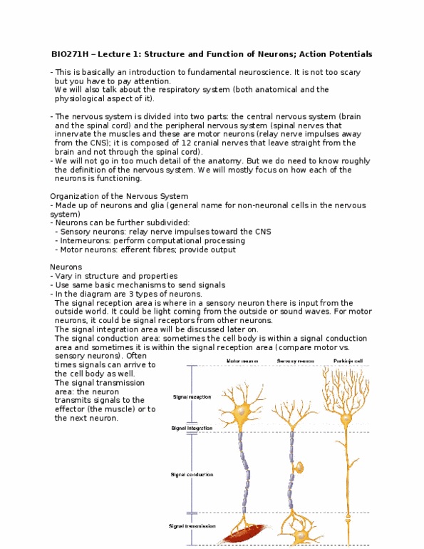
Action potential arrives in pre-synaptic cell1)
Acetylcholine (Ach) is released into synaptic cleft2)
Opening of Na-channels and generation of post-synaptic action
potential
3)
Propagation of electrical signal in post-synaptic cell (muscle) 4)
Neuromuscular Junction
Brain and nerves within cell body (soma) in brain case or
spinal cord
○
Central Nervous System
!
Nerves will cell body outside brain case or spinal cord
○
Peripheral Nervous System
!
Organization of the Nervous System
Membrane potential -difference in charge between inside
and outside cell
○
Caused by differences in ion concentrations across cell
membrane
○
Neurons have a resting membrane potential (like all cells)
!
Neurons are excitable
!
*see slide
○
Resting membrane potential (-70 mV)
○
Depolarization (increases to -60 mV)
○
Repolarization (decreases to -70 mV)
○
Hyperpolarization (decreases to -75 mV)
○
Repolarization (increases to -70 mV)
○
Changes in membrane potential acts as electrical signals
!
The voltage difference seen across cell membranes is due to the
movement of ions (distribution across the membrane)
!
Electrical Signals in Neurons
Chemical gradient
○
Electrical gradient (electromotive force)
○
The distribution of an ion across an ion-selective membrane
depends on two opposing forces:
!
K+ equilibrium potential = EK
○
The potential difference across the membrane when the two
forces are in equilibrium = equilibrium potential
!
EX= RT/zF ln( [X]out / [X]in)
!
R = gas constant
!
T = temperature
!
z = valence of ion
!
F = faraday constant
!
EXis proportional to the ratio of the concentrations of ion
X across the membrane
○
Ex(V) = 0.058/z log( [X]out / [X]in)
!
Ex(mV) = 58/z log( [X]out / [X]in)
!
For example, at 18 degrees:
○
[Na+] -10mM in, 120 mM out
"
[K+] -140mM in, 2.5 mM out
"
[Ca2+] -0.001mM in, 2mM out
"
[Cl-] -4mM in, 120 mM out
"
Assuming the following distribution of ions across a
cell membrane:
!
ENa+ = 58/1 log(120 / 10) = +63 mV (depolarize)
!
EK+ = 58/1 log(2.5/140) = -101 mV (hyperpolarize)
!
ECl- = 58/-1 log(120/4) = -86 mV (hyperpolarize)
!
ECa2+ = 58/2 log(2/0.001) = +96mV (depolarize)
!
Ex:
○
Nernst Equation: used to calculate the equilibrium potential for
single ions
!
Vm= RT/F * ln [ (PK[K+]o+ PNa[Na+]o+ PCl-[Cl-]i
+ Pca[Ca2+]o) / (PK[K+]i+ PNa[Na+]i+ PCl-[Cl-]o+
Pca[Ca2+]i)]
!
P = permeability
!
R = gas constant
!
T = absolute temperature (K)
!
F = Faraday constant
!
Vmis proportional to the ratio of the ionic membrane
concentrations across the membrane and the permeability
of the membrane to ions
○
Vm= RT/F * ln [ (1[K+]o+ 0.01[Na+]o) / (1[K+]i+
0.01[Na+]i) ]
!
Since all cell membranes have very low permeability to
Ca2+ and Cl-, and the permeability to Na+ is about 1/100
that of K+, the equation can be simplified to:
○
Vm= 58* log [ (2.5 + (0.01*120) / (140 + (0.01*
10) ] = -92mV
!
Therefore, at 18 degrees:
○
Goldman Equation: used to calculate the final membrane
potential (Vm) from all the contributing ions
!
Cell membranes have a higher permeability to K+
than to other ions
!
The Na+/K+ pump indirectly contributes to Vmby
maintaining the high internal [K+]
!
The Vmof a cell (-92 mV) is relatively close to EK(-101
mV) because:
○
The role of K+ and the Na+/K+ Pump for Resting Membrane
Potential
!
Electrochemical Gradients and Membrane Potentials
Structure-function relationships of neurons
!
Ligand-gated ion channels
○
Properties of graded potentials
○
Signals in the dendrites and soma
!
Properties of action potentials
○
Signals in the axon
!
Outline:
Readings: Ch. 12 (309-320)
Stimulus -sensory neuron -giant interneuron -leg
motor neuron -muscle tension
!
Ex. Neural circuit mediating the startle response in
cockroach
○
Neurons are organized into functional circuits that rapidly
conduct information to a target
!
Signal reception (input) -dendrites
○
Signal integration -axon hillock
○
Signal conduction -axon (--> all-or-none response of
action potential)
○
Signal transmission (output) -axon terminals
○
Four functional zones:
!
Information is carried through neuronal circuits via
alternating electrical signals and chemical signals
○
Sensory neurons are afferent fibers and carry information
inward toward interneurons
○
Motor neurons (interneuron) are efferent fibers and carry
information outward to effectors (like muscles)
○
Information through neuronal circuits alternate between graded
and all-or-none signals
!
Structure-Function Relationships
Ligand-gated ion channels convert chemical signals into
electrical signals by changing the membrane potential
!
Nicotinic acethycholine receptors
○
Glutamate receptors
○
GABAAreceptors
○
Examples:
!
In the dendrites and soma the electrical signals generated by
ligant-gated ion channels are called graded potentials
!
No neurotransmitter --> ion channel is closed (no
crossing of ions)
!
Low [neurotransmitter] --> some ions can cross
membrane
!
High [neurotransmitter] --> many ions can cross
membrane (neurotransmitters are bound to most
receptors)
!
The magnitude is proportional to the stimulus strength (to
the concentration of neurotransmitter)
○
Ex. ENa+ = 58/1 * log(120/10) = +63mV --> depolarizes
membrane
○
Graded potentials vary in magnitude:
!
Depolarize the cell (Na+ and Ca2+)
○
Hyperpolarize the cell (K+ and Cl-)
○
Graded potentials can:
!
Net movement stops when the equilibrium potential is
reached
○
Ions move down an electrochemical gradient
!
Membrane permeability -leakage of charged ions
across the membrane
!
Cytoplasmic resistance -inherent resistance to
current flow
!
Due to:
○
The decremental spread of graded potentials = electrotonic
conduction
○
Neurotransmitter binds to ligand-gated Na+ channel
!
Na+ enters cell through open channel
!
Current spreads through the cell
!
The signal of the strength decreases with distance
!
Ex.
○
Graded potentials are short-distance signals:
!
Sub-threshold graded potentials do not initiate an
action potential
!
Supra-threshold graded potentials cause the axon to
fire an action potential
!
Graded potentials in the axon hillock need to depolarize
the membrane beyond the threshold potential in order for
the axon to fire an action potential
○
Transition from graded to all-or-none response:
!
Signals in the Dendrites and Soma
Spatial summation: graded potentials from different locations
can interact to influence the net change in membrane potential at
the axon hillock
!
Temporal summation: graded potentials occurring at slightly
different times can interact to influence the net graded potential
!
Sub-threshold potential that overlap in time summate and
may trigger an action potential
○
Sub-threshold potentials that do not overlap in time do not
summate
!
Graded potentials are integrated to trigger action potentials
Stimulating current pulses -hyperpolarizing current
(decrease potential) -depolarizing current (increase
potential)
○
*see slide
!
All supra-threshold stimulus produce an identical action
potential
!
Relationship between Stimulus and Response
Triggered when membrane potential at axon hillock reaches
threshold
!
Large (~100mV), brief (2-3msec) propagated change in Vm
!
Once triggered, AP is an all-or-none response
!
Current is carried by ions (not electrons)
!
AP formation does not require ATP
!
Ion concentrations are restored by Na+/K+ ATPase pump
!
Depolarizing graded potential
○
Depolarization phase of action potential -absolute
refractory period
○
Repolarization phase of action potential
○
Hyperpolarization -relative refractory period (return to
resting membrane potential)
○
*see slide
!
Properties of Action Potentials
Voltage-gated Na+ and K+ channels
!
Propagation of action potentials
!
Ionic basis of the action potential
○
Signals in the axon
!
The length constant
○
The time constant
○
The importance of axon diameter and myelination
○
Factors affecting the neuronal conduction speed
!
Outline:
Readings: Chapter 12 (pages 320-325)
See slide
!
Uses glass dish containing isolated neurons and an electrode
!
Single patch electrode is useful to examine neurons
!
Uses suction to remove membrane patch
!
Pipette solution composition = extracellular
!
Tissue bath solution composition = intracellular
!
Micropipette electrode suctions up part of membrane with
voltage-gated Na+ channel
○
One can determine how the voltage and current flowing
through cell changes when the Na+ channel is open (when
part of membrane is removed)
○
Each Na+ channel opens with little delay following initial
depolarization and stays open for less than a millisecond
before becoming inactivated
○
The voltage-gated K+ channels open slightly later and can
stay open until shortly after membrane repolarization
○
Patch-clamp recording of single-channel currents:
!
Single Cell Patch Clamp Rig
Rising -falling -after-hyperpolarization
○
Opening and closing of voltage-gated ion channels cause the
characteristic phases of the action potential
!
Depolarization involves a 30x increase in Na+ conductance (gNa)
!
Repolarization involves a decrease in gNa and a delayed increase
in gK
!
After-hyperpolarization occurs because gKremains elevated for
some time after the action potential
!
Ionic Basis of the Action Potential
Contains two gates, a voltage-dependent activation gate
and a voltage-dependent time-delayed inactivation gate
○
Closed but capable of opening -at resting potential
(-70 mV)
!
Open and activated -from threshold to peak
potential (-50 to +30 mV)
!
Closed and not capable of opening aka inactivated -
from peak to resting potential (+30 mV to -50 mV)
!
The voltage-gated Na+ channel can exist in 3 different
conformations:
○
Na+ Channels:
!
At resting potential
"
Delayed opening triggered at threshold
"
Remains closed to peak potential (-70 to +30
mV)
"
Closed:
!
From peak potential through after
hyperpolarization (+30 to -80 mV)
"
Open:
!
Voltage-gated K+ channel has only one voltage-dependent
time-delayed gate that can either be open or closed
○
K+ Channels:
!
Voltage-Gated Channels
Action potentials move down the axon without decrement
!
Na+ local currents spread longitudinally (via electrotonic
conduction), depolarizing adjacent patches
!
The inactivation gate prevents action potentials from travelling
backwards
!
Na+ ions move through voltage-gated channel
○
Current flows through activated patch of membrane and
depolarizes the adjacent patch
○
*repolarized patch is refractory, so action potential
travels in one direction
!
Adjacent patch reaches threshold, current flows and
depolarizes next adjacent patch
○
*after refractory period, it is ready to be activated
again
!
Process continues
○
Steps:
!
Propagation of Action Potentials
After an action potential is triggered, neurons enter a refractor
period
!
No action potential can be triggered during the absolute
refractory period (why?)
!
It is more difficult to generate a new action potential during the
relative refractory period (why?)
!
What are the advantages of the refractory period?
!
*see slide
!
Properties of Action Potentials
Passive spread (electrotonic)
○
Action potentials
○
Saltatory conduction
○
Chemical and eletrical synapses
○
Signal conduction can be via:
!
Is a combination of electrotonic flow and action potentials
○
But electrotonic current flow is graded and can only
travel short distances
!
Electrotonic current flow is much faster than action
potentials
○
Axonal conduction:
!
Diversity of Signal Conduction
Electrotonic conduction is enhanced by high membrane
resistance and low longitudinal (axoplasmic) resistance
○
The decay of Vm with distance is described by the length
constant: λ
○
λ = sq. rt (Rm / Rl)
!
Rm = membrane resistance
"
Rl = longitudinal resistance
"
Where…
!
λis defined as the distance over which Vm falls by 63%
of its initial value
○
The length Constant (λ)
1)
Membrane voltage changes are reduced by high
membrane capacitance and resistance
○
Following an applied voltage, the time needed to reach a
given Vm is described by the time constant: τ
○
τ = Rm * Cm
!
Rm = membrane resistance
"
Cm = membrane capacitance
"
Where…
!
τis defined as the time taken for Vm to reach 63% of its
maximal value
○
The time Constant (τ)
2)
Factors Affecting Conduction Speed
E.g. fatty membranes of glial cells:
olgiodendrocytes or Schwann cells
!
The axon of some neurons are wrapped with myelin
○
Myelination greatly increases the length constant (why?)
○
Segmented myelination leads to fast saltatory conduction
of action potentials (how?)
○
Overall, myelinated axons speed the propagation of an
action potential
○
*see slide
!
Somatodendritic input = synapse
!
Axonal output = axon initial segment -nodes of
ranvier (-neuromuscular junction)
!
Spatial distribution of voltage-gated channels at the
surface of a myelinated neuron:
○
Axon Myelination1)
Increasing axonal diameter increases the length constant
and conduction velocity (why?)
Myelinated fibers have a larger axon diameter and
conduction velocity
!
Each vertebrate nerve contains a mixture of different
neuronal fibre types
○
Conduction velocity increases with axon diameter
across species
!
*see slide
○
Axon Diameter2)
Factors Affecting Speed of Propagation
Electrical synapse
○
Fast vs. slow c
!
Chemical synapse
○
Signals across the synapse
!
Structural specializations
○
Events at a neuromuscular junction
○
Acetylcholine
○
Signals across neuromuscular junction
!
Outline:
Presynaptic cell
○
Synaptic cleft
○
Postsynaptic cell
○
A signal transmission zone consisting of:
!
Synaptic cleft -space between pre and postsynaptic cell
!
Postsynaptic cell -neurons, muscles, and endocrine glands
!
Neuromuscular junction -synapse between a motor neuron and
a muscle
!
The Synapse
Electrical synapses transfer information between cells by direct
ionic coupling via gap junction
!
Current decays between neurons (just like passive spread of
local Na+ current)
!
The connexon proteins of gap junctions narrow the jap and
lower the resistance between cells
!
Advantage -very rapid
○
Disadvantage -requires diffusion from connection;
weakens with distance
○
What is the principle advantage and disadvantage of electrical
synapses?
!
*see current flow at electrical vs chemical synapses
!
*see electrical synapses in the crayfish escape circuit
○
Ventral nerve cord -giant nerve ganglion
○
Electrical synapses were first demonstrated between ventral
nerve cord giant axons and the motor neuros responsible for the
tail-flip escape response of crayfish
!
Electrical Synapses
Presynaptic terminal -synaptic vesicles (AcH) -
presynaptic densities -synaptic cleft -postsynaptic
densities
○
Dendrite -(synapse) -dendritic spines
○
1 mitochondria per bouton --> energetically expensive
○
*see structure
!
Chemical signals transfer information between cells indirectly
via neurotransmitters
!
The amount of neurotransmitter released is influenced by
intracellular Ca2+ which is influenced by AP frequency
and by mechanisms that regulate [Ca2+]
○
Intracellular Ca2+ regulates neurotransmitter release
!
Fast and slow chemical synapses are defined by their post-
synaptic mechanisms (not their neurotransmitters)
○
Fast chemical synapses act through ionotropic receptors
(i.e. ligand-gated ion channels) on the post synaptic
membrane
○
Slow chemical synapses act through metabotropic
receptors on the post synaptic membrane
○
*see mechanism figures on slides
○
Fast vs. Slow Synaptic Transmission:
!
Chemical Synapses
Electrical Chemical
Rare in complex animals Common in complex animals
Comparatively fast Comparatively slow
Bi-directional Unidirectional
Postsynaptic signal is similar to
presynaptic
(weakens but it is still the same
electrical signal)
Postsynaptic signal can be
different
Excitatory Excitatory (Na+) or
inhibitory (K+)
Action potentials are conducted to skeletal muscles through
large, myelinated motor neurons
!
Pre-synaptic terminal boutons (for significant SA)
○
Schwann-cell sheath (provide insulation so there is no loss
of signal)
○
Basement membrane
○
Junctional folds (greater SA)
○
The neuromuscular junction includes pre and postsynaptic
specializations
!
Presynaptic action potential reaches pre-synaptic cell
causing influx of Ca2+
○
Changes in Na+ (in) and K+ (out)
!
This results in Ach exocytosis from pre-synaptic cell
(from vesicles) and action of Ach on postsynaptic receptor
○
Current causes end plate potential --> postsynaptic action
potential
○
*steps (getting rid of ACh quickly) allows for another
action potential to occur quickly
○
Events at a neuromuscular junction (chemical synapse):
!
Primary neurotransmitter at the vertebrate neuromuscular
junction
○
Synthesis and recycling of ACh occurs at the synapse
○
Neurotransmitter amount: rate of release vs. rate of
removal
!
Receptor activity: density of receptors on
postsynaptic cells
!
Signal strength is influenced by neurotransmitter amount
and receptor activity
○
Acetylcholine (ACh):
!
The Neuromuscular Junction: Structural Specializations
What is neuronal/synaptic plasticity?
!
Plasticity is rooted in diversity at the chemical synapse
!
Habituation and sensitization in Aplysia
○
Example of short-term neuromodulation:
!
Potential in hippocampal neurons
○
Example of long-term neuromodulation:
!
Outline:
EPSPs move Vm toward threshold potential
!
IPSPs move Vm away from threshold potential
!
*note: smaller distance from electrode = less resistance
(electrotonic properties)
○
EPSPs and IPSPs summate (temporal and spatial)
!
Excitatory and Inhibitory Postsynaptic Potentials
Plasticity -ability to change synaptic strength over time via both
synaptic connections and functional properties of neurons
!
The synaptic transfer of information depends on its history
!
Facilitation -strength of response increases over time (change in
charge increases)
!
What are the mechanisms?
○
Learning -process of acquiring new information
!
What are the mechanisms?
○
Memory -retention and retrieval of information
!
To make connection stronger: more proteins, more ACh …etc
(in chemical synapse)
!
Neuronal Plasticity
Biogenic amines
○
Amino acids
○
Neuropeptides
○
Others
○
Chemical synapses use many types of neurotransmitters and
receptors:
!
Ex. Ionotropic Ach nicotinic receptor (faster?)
○
Ex. Metabotropic ACh muscarinic receptor
○
Whether a neurotransmitter is excitatory or inhibitory depends
on the properties of its receptors
!
Many neurons can synthesize more than one kind of
neurotransmitter
!
Plasticity is rooted in diversity at the chemical synapse
Stimulation of the mantle or siphon leads to gill
withdrawal (escape response)
○
Over time, the amplitude of withdrawal decreases =
learning response
!
This reflex response habituates with repeated stimulation
(gets used to stimulus so it doesn't respond)
○
After tapping on head, tapping on the siphon
causing gill withdrawal (after being habituated)
!
This reflex response can also be enhanced (sensitized) in
response to a novel stimulus (i..e. tapping on the head)
○
The reduction and enhancement of the motor-
neuron excitatory post synaptic potentials mirror the
behavioural habituation and sensitization,
respectively
!
Sensitization involves a secondary facilitating
interneuron
!
The gill-withdrawal reflex can be studies at the synaptic
level and at the whole animal level
○
Ex. Gill-withdrawal reflex in sea slug (Aplysia)
!
Serotonin --> serotonin receptor --> G protein -->
cAMP
!
cAMP-dependent kinase acts on voltage-gated K+
(via phosphorylation) --> slows down rate of K+
leaving cell so Ca2+ can enter cell for a longer
amount of time (so more neurotransmitters are
released via activation of vesicles by Ca)
!
Causes more proteins being activated by the
neurotransmitter to increase strength of action
potential
!
Short-term sensitization occurs from a increase in
neurotransmitter release as a result of presynaptic
facilitation
○
cAMP can cause nucleus to increase number of
channels
!
Note: Long-term sensitization can occur if kinase activity
elicits changes in sensory neuron protein synthesis (from
repeated trials of the novel stimulus)
○
Short term-habituation occurs from a reduction in
neurotransmitter release by the sensory neuron (activating less
ligand-gated channels; decrease in Ca2+ entering neuron)
!
Short-term neuromodulation: habituation & sensitization
How? -via changes in post-synaptic cell (stronger
depolarization)
!
Tetanic stimulation of neurons (10 pps for 10s;
represented as learning) in the hippocampus leads to an
increase in EPSPs
○
NMDA receptors are blocked at resting potential by
Mg2+ ions
!
Lets Na+ in and K+ out
"
AMPA receptors open to produce a fast EPSP
!
Ca+ in, K+ out, and N+ in
!
Ca2+ ions enter the NMDA receptor
channels in the post synaptic cell and
activate Ca2+ dependent protein kinases
!
Depolarized Mg2+ (with Glutamate binding)
so ions can flow through NMDA
"
This fusion delivers new receptors and
new lipid membrane to the spine
head --> allows a stronger signal
!
Ca2+ triggered phosphorylation of AMPA
receptors stored in internal vesicles stimulates
fusion of the vesicles with the cell membrane
(over time will make more proteins due to
increase in rates of transcription and
translation)
"
With 100pps stimulation:
!
*this is how learning works (connections made are
strong; and repetition increases strength over time)
!
Induction and maintenance of LTP in the hippocampus:
○
Ex. Long-term potentiation (LTP) in the mammalian
hippocampus
!
Long-term neuromodulation: potentiation
Glutamate release -CaMKII (Ca2+ activated kinase) -
single dendritic spine increases (due to increase in
membrane --> larger SA)
○
Increase in volume of the dendritic spine:
!
Effect of increased glutamate on a single dendritic spine:
Neurons
#$%&'()*+, -./0.12.&, 34+,3546
4758,9:

Structure-Function Relationship of Neurons
!
Glial Cells
!
Electrical Signals in Neurons
!
The Nernst Equation
!
The Goldman Equation
!
Electrochemical potentials
○
Membrane Potential -mathematical underpinnings
!
Outline:
Readings: Chapter 12 (pages 295-308), Invertebrate Studies and their
Contribution to Neuroscience
Neuron -anatomical and functional unit of the nervous system
Signal reception (input): dendrites
○
Signal integration: axon hillock
○
Signal conduction: axon
○
Signal transmission (output): axon terminals
○
Four functional zones:
!
Synapse: connection between two nerves or a nerve cell and
muscle cell
!
Cell body (soma) to axon terminals
○
Neural signals only travel in one direction
!
MS patients lose their myelin sheaths which causes signal
strength to decrease and other complications
○
Myelin sheath insulates the neuron
!
Structure-Function Relationship of Neurons
Most neural cells are glial cells
!
Glial cells support neurons
!
They do not generate action potentials
!
Schwann cell: form myelin in motor and sensory neurons
of the PNS
○
Olgiodendrocyte: form myelin in the CNS
○
Astrocyte: transport nutrients, remove debris in CNS,
regulate synaptic neurotransmitter signals
○
Microglia: remove debris and dead cells from CNS
○
Four main types:
!
Glial Cells
Action potential arrives in pre-synaptic cell
1)
Acetylcholine (Ach) is released into synaptic cleft
2)
Opening of Na-channels and generation of post-synaptic action
potential
3)
Propagation of electrical signal in post-synaptic cell (muscle)
4)
Neuromuscular Junction
Brain and nerves within cell body (soma) in brain case or
spinal cord
○
Central Nervous System
!
Nerves will cell body outside brain case or spinal cord
○
Peripheral Nervous System
!
Organization of the Nervous System
Membrane potential -difference in charge between inside
and outside cell
○
Caused by differences in ion concentrations across cell
membrane
○
Neurons have a resting membrane potential (like all cells)
!
Neurons are excitable
!
*see slide
○
Resting membrane potential (-70 mV)
○
Depolarization (increases to -60 mV)
○
Repolarization (decreases to -70 mV)
○
Hyperpolarization (decreases to -75 mV)
○
Repolarization (increases to -70 mV)
○
Changes in membrane potential acts as electrical signals
!
The voltage difference seen across cell membranes is due to the
movement of ions (distribution across the membrane)
!
Electrical Signals in Neurons
Chemical gradient
○
Electrical gradient (electromotive force)
○
The distribution of an ion across an ion-selective membrane
depends on two opposing forces:
!
K+ equilibrium potential = EK
○
The potential difference across the membrane when the two
forces are in equilibrium = equilibrium potential
!
EX= RT/zF ln( [X]out / [X]in)
!
R = gas constant
!
T = temperature
!
z = valence of ion
!
F = faraday constant
!
EXis proportional to the ratio of the concentrations of ion
X across the membrane
○
Ex(V) = 0.058/z log( [X]out / [X]in)
!
Ex(mV) = 58/z log( [X]out / [X]in)
!
For example, at 18 degrees:
○
[Na+] -10mM in, 120 mM out
"
[K+] -140mM in, 2.5 mM out
"
[Ca2+] -0.001mM in, 2mM out
"
[Cl-] -4mM in, 120 mM out
"
Assuming the following distribution of ions across a
cell membrane:
!
ENa+ = 58/1 log(120 / 10) = +63 mV (depolarize)
!
EK+ = 58/1 log(2.5/140) = -101 mV (hyperpolarize)
!
ECl- = 58/-1 log(120/4) = -86 mV (hyperpolarize)
!
ECa2+ = 58/2 log(2/0.001) = +96mV (depolarize)
!
Ex:
○
Nernst Equation: used to calculate the equilibrium potential for
single ions
!
Vm= RT/F * ln [ (PK[K+]o+ PNa[Na+]o+ PCl-[Cl-]i
+ Pca[Ca2+]o) / (PK[K+]i+ PNa[Na+]i+ PCl-[Cl-]o+
Pca[Ca2+]i)]
!
P = permeability
!
R = gas constant
!
T = absolute temperature (K)
!
F = Faraday constant
!
Vmis proportional to the ratio of the ionic membrane
concentrations across the membrane and the permeability
of the membrane to ions
○
Vm= RT/F * ln [ (1[K+]o+ 0.01[Na+]o) / (1[K+]i+
0.01[Na+]i) ]
!
Since all cell membranes have very low permeability to
Ca2+ and Cl-, and the permeability to Na+ is about 1/100
that of K+, the equation can be simplified to:
○
Vm= 58* log [ (2.5 + (0.01*120) / (140 + (0.01*
10) ] = -92mV
!
Therefore, at 18 degrees:
○
Goldman Equation: used to calculate the final membrane
potential (Vm) from all the contributing ions
!
Cell membranes have a higher permeability to K+
than to other ions
!
The Na+/K+ pump indirectly contributes to Vmby
maintaining the high internal [K+]
!
The Vmof a cell (-92 mV) is relatively close to EK(-101
mV) because:
○
The role of K+ and the Na+/K+ Pump for Resting Membrane
Potential
!
Electrochemical Gradients and Membrane Potentials
Structure-function relationships of neurons
!
Ligand-gated ion channels
○
Properties of graded potentials
○
Signals in the dendrites and soma
!
Properties of action potentials
○
Signals in the axon
!
Outline:
Readings: Ch. 12 (309-320)
Stimulus -sensory neuron -giant interneuron -leg
motor neuron -muscle tension
!
Ex. Neural circuit mediating the startle response in
cockroach
○
Neurons are organized into functional circuits that rapidly
conduct information to a target
!
Signal reception (input) -dendrites
○
Signal integration -axon hillock
○
Signal conduction -axon (--> all-or-none response of
action potential)
○
Signal transmission (output) -axon terminals
○
Four functional zones:
!
Information is carried through neuronal circuits via
alternating electrical signals and chemical signals
○
Sensory neurons are afferent fibers and carry information
inward toward interneurons
○
Motor neurons (interneuron) are efferent fibers and carry
information outward to effectors (like muscles)
○
Information through neuronal circuits alternate between graded
and all-or-none signals
!
Structure-Function Relationships
Ligand-gated ion channels convert chemical signals into
electrical signals by changing the membrane potential
!
Nicotinic acethycholine receptors
○
Glutamate receptors
○
GABAAreceptors
○
Examples:
!
In the dendrites and soma the electrical signals generated by
ligant-gated ion channels are called graded potentials
!
No neurotransmitter --> ion channel is closed (no
crossing of ions)
!
Low [neurotransmitter] --> some ions can cross
membrane
!
High [neurotransmitter] --> many ions can cross
membrane (neurotransmitters are bound to most
receptors)
!
The magnitude is proportional to the stimulus strength (to
the concentration of neurotransmitter)
○
Ex. ENa+ = 58/1 * log(120/10) = +63mV --> depolarizes
membrane
○
Graded potentials vary in magnitude:
!
Depolarize the cell (Na+ and Ca2+)
○
Hyperpolarize the cell (K+ and Cl-)
○
Graded potentials can:
!
Net movement stops when the equilibrium potential is
reached
○
Ions move down an electrochemical gradient
!
Membrane permeability -leakage of charged ions
across the membrane
!
Cytoplasmic resistance -inherent resistance to
current flow
!
Due to:
○
The decremental spread of graded potentials = electrotonic
conduction
○
Neurotransmitter binds to ligand-gated Na+ channel
!
Na+ enters cell through open channel
!
Current spreads through the cell
!
The signal of the strength decreases with distance
!
Ex.
○
Graded potentials are short-distance signals:
!
Sub-threshold graded potentials do not initiate an
action potential
!
Supra-threshold graded potentials cause the axon to
fire an action potential
!
Graded potentials in the axon hillock need to depolarize
the membrane beyond the threshold potential in order for
the axon to fire an action potential
○
Transition from graded to all-or-none response:
!
Signals in the Dendrites and Soma
Spatial summation: graded potentials from different locations
can interact to influence the net change in membrane potential at
the axon hillock
!
Temporal summation: graded potentials occurring at slightly
different times can interact to influence the net graded potential
!
Sub-threshold potential that overlap in time summate and
may trigger an action potential
○
Sub-threshold potentials that do not overlap in time do not
summate
!
Graded potentials are integrated to trigger action potentials
Stimulating current pulses -hyperpolarizing current
(decrease potential) -depolarizing current (increase
potential)
○
*see slide
!
All supra-threshold stimulus produce an identical action
potential
!
Relationship between Stimulus and Response
Triggered when membrane potential at axon hillock reaches
threshold
!
Large (~100mV), brief (2-3msec) propagated change in Vm
!
Once triggered, AP is an all-or-none response
!
Current is carried by ions (not electrons)
!
AP formation does not require ATP
!
Ion concentrations are restored by Na+/K+ ATPase pump
!
Depolarizing graded potential
○
Depolarization phase of action potential -absolute
refractory period
○
Repolarization phase of action potential
○
Hyperpolarization -relative refractory period (return to
resting membrane potential)
○
*see slide
!
Properties of Action Potentials
Voltage-gated Na+ and K+ channels
!
Propagation of action potentials
!
Ionic basis of the action potential
○
Signals in the axon
!
The length constant
○
The time constant
○
The importance of axon diameter and myelination
○
Factors affecting the neuronal conduction speed
!
Outline:
Readings: Chapter 12 (pages 320-325)
See slide
!
Uses glass dish containing isolated neurons and an electrode
!
Single patch electrode is useful to examine neurons
!
Uses suction to remove membrane patch
!
Pipette solution composition = extracellular
!
Tissue bath solution composition = intracellular
!
Micropipette electrode suctions up part of membrane with
voltage-gated Na+ channel
○
One can determine how the voltage and current flowing
through cell changes when the Na+ channel is open (when
part of membrane is removed)
○
Each Na+ channel opens with little delay following initial
depolarization and stays open for less than a millisecond
before becoming inactivated
○
The voltage-gated K+ channels open slightly later and can
stay open until shortly after membrane repolarization
○
Patch-clamp recording of single-channel currents:
!
Single Cell Patch Clamp Rig
Rising -falling -after-hyperpolarization
○
Opening and closing of voltage-gated ion channels cause the
characteristic phases of the action potential
!
Depolarization involves a 30x increase in Na+ conductance (gNa)
!
Repolarization involves a decrease in gNa and a delayed increase
in gK
!
After-hyperpolarization occurs because gKremains elevated for
some time after the action potential
!
Ionic Basis of the Action Potential
Contains two gates, a voltage-dependent activation gate
and a voltage-dependent time-delayed inactivation gate
○
Closed but capable of opening -at resting potential
(-70 mV)
!
Open and activated -from threshold to peak
potential (-50 to +30 mV)
!
Closed and not capable of opening aka inactivated -
from peak to resting potential (+30 mV to -50 mV)
!
The voltage-gated Na+ channel can exist in 3 different
conformations:
○
Na+ Channels:
!
At resting potential
"
Delayed opening triggered at threshold
"
Remains closed to peak potential (-70 to +30
mV)
"
Closed:
!
From peak potential through after
hyperpolarization (+30 to -80 mV)
"
Open:
!
Voltage-gated K+ channel has only one voltage-dependent
time-delayed gate that can either be open or closed
○
K+ Channels:
!
Voltage-Gated Channels
Action potentials move down the axon without decrement
!
Na+ local currents spread longitudinally (via electrotonic
conduction), depolarizing adjacent patches
!
The inactivation gate prevents action potentials from travelling
backwards
!
Na+ ions move through voltage-gated channel
○
Current flows through activated patch of membrane and
depolarizes the adjacent patch
○
*repolarized patch is refractory, so action potential
travels in one direction
!
Adjacent patch reaches threshold, current flows and
depolarizes next adjacent patch
○
*after refractory period, it is ready to be activated
again
!
Process continues
○
Steps:
!
Propagation of Action Potentials
After an action potential is triggered, neurons enter a refractor
period
!
No action potential can be triggered during the absolute
refractory period (why?)
!
It is more difficult to generate a new action potential during the
relative refractory period (why?)
!
What are the advantages of the refractory period?
!
*see slide
!
Properties of Action Potentials
Passive spread (electrotonic)
○
Action potentials
○
Saltatory conduction
○
Chemical and eletrical synapses
○
Signal conduction can be via:
!
Is a combination of electrotonic flow and action potentials
○
But electrotonic current flow is graded and can only
travel short distances
!
Electrotonic current flow is much faster than action
potentials
○
Axonal conduction:
!
Diversity of Signal Conduction
Electrotonic conduction is enhanced by high membrane
resistance and low longitudinal (axoplasmic) resistance
○
The decay of Vm with distance is described by the length
constant: λ
○
λ = sq. rt (Rm / Rl)
!
Rm = membrane resistance
"
Rl = longitudinal resistance
"
Where…
!
λis defined as the distance over which Vm falls by 63%
of its initial value
○
The length Constant (λ)
1)
Membrane voltage changes are reduced by high
membrane capacitance and resistance
○
Following an applied voltage, the time needed to reach a
given Vm is described by the time constant: τ
○
τ = Rm * Cm
!
Rm = membrane resistance
"
Cm = membrane capacitance
"
Where…
!
τis defined as the time taken for Vm to reach 63% of its
maximal value
○
The time Constant (τ)
2)
Factors Affecting Conduction Speed
E.g. fatty membranes of glial cells:
olgiodendrocytes or Schwann cells
!
The axon of some neurons are wrapped with myelin
○
Myelination greatly increases the length constant (why?)
○
Segmented myelination leads to fast saltatory conduction
of action potentials (how?)
○
Overall, myelinated axons speed the propagation of an
action potential
○
*see slide
!
Somatodendritic input = synapse
!
Axonal output = axon initial segment -nodes of
ranvier (-neuromuscular junction)
!
Spatial distribution of voltage-gated channels at the
surface of a myelinated neuron:
○
Axon Myelination1)
Increasing axonal diameter increases the length constant
and conduction velocity (why?)
Myelinated fibers have a larger axon diameter and
conduction velocity
!
Each vertebrate nerve contains a mixture of different
neuronal fibre types
○
Conduction velocity increases with axon diameter
across species
!
*see slide
○
Axon Diameter2)
Factors Affecting Speed of Propagation
Electrical synapse
○
Fast vs. slow c
!
Chemical synapse
○
Signals across the synapse
!
Structural specializations
○
Events at a neuromuscular junction
○
Acetylcholine
○
Signals across neuromuscular junction
!
Outline:
Presynaptic cell
○
Synaptic cleft
○
Postsynaptic cell
○
A signal transmission zone consisting of:
!
Synaptic cleft -space between pre and postsynaptic cell
!
Postsynaptic cell -neurons, muscles, and endocrine glands
!
Neuromuscular junction -synapse between a motor neuron and
a muscle
!
The Synapse
Electrical synapses transfer information between cells by direct
ionic coupling via gap junction
!
Current decays between neurons (just like passive spread of
local Na+ current)
!
The connexon proteins of gap junctions narrow the jap and
lower the resistance between cells
!
Advantage -very rapid
○
Disadvantage -requires diffusion from connection;
weakens with distance
○
What is the principle advantage and disadvantage of electrical
synapses?
!
*see current flow at electrical vs chemical synapses
!
*see electrical synapses in the crayfish escape circuit
○
Ventral nerve cord -giant nerve ganglion
○
Electrical synapses were first demonstrated between ventral
nerve cord giant axons and the motor neuros responsible for the
tail-flip escape response of crayfish
!
Electrical Synapses
Presynaptic terminal -synaptic vesicles (AcH) -
presynaptic densities -synaptic cleft -postsynaptic
densities
○
Dendrite -(synapse) -dendritic spines
○
1 mitochondria per bouton --> energetically expensive
○
*see structure
!
Chemical signals transfer information between cells indirectly
via neurotransmitters
!
The amount of neurotransmitter released is influenced by
intracellular Ca2+ which is influenced by AP frequency
and by mechanisms that regulate [Ca2+]
○
Intracellular Ca2+ regulates neurotransmitter release
!
Fast and slow chemical synapses are defined by their post-
synaptic mechanisms (not their neurotransmitters)
○
Fast chemical synapses act through ionotropic receptors
(i.e. ligand-gated ion channels) on the post synaptic
membrane
○
Slow chemical synapses act through metabotropic
receptors on the post synaptic membrane
○
*see mechanism figures on slides
○
Fast vs. Slow Synaptic Transmission:
!
Chemical Synapses
Electrical Chemical
Rare in complex animals Common in complex animals
Comparatively fast Comparatively slow
Bi-directional Unidirectional
Postsynaptic signal is similar to
presynaptic
(weakens but it is still the same
electrical signal)
Postsynaptic signal can be
different
Excitatory Excitatory (Na+) or
inhibitory (K+)
Action potentials are conducted to skeletal muscles through
large, myelinated motor neurons
!
Pre-synaptic terminal boutons (for significant SA)
○
Schwann-cell sheath (provide insulation so there is no loss
of signal)
○
Basement membrane
○
Junctional folds (greater SA)
○
The neuromuscular junction includes pre and postsynaptic
specializations
!
Presynaptic action potential reaches pre-synaptic cell
causing influx of Ca2+
○
Changes in Na+ (in) and K+ (out)
!
This results in Ach exocytosis from pre-synaptic cell
(from vesicles) and action of Ach on postsynaptic receptor
○
Current causes end plate potential --> postsynaptic action
potential
○
*steps (getting rid of ACh quickly) allows for another
action potential to occur quickly
○
Events at a neuromuscular junction (chemical synapse):
!
Primary neurotransmitter at the vertebrate neuromuscular
junction
○
Synthesis and recycling of ACh occurs at the synapse
○
Neurotransmitter amount: rate of release vs. rate of
removal
!
Receptor activity: density of receptors on
postsynaptic cells
!
Signal strength is influenced by neurotransmitter amount
and receptor activity
○
Acetylcholine (ACh):
!
The Neuromuscular Junction: Structural Specializations
What is neuronal/synaptic plasticity?
!
Plasticity is rooted in diversity at the chemical synapse
!
Habituation and sensitization in Aplysia
○
Example of short-term neuromodulation:
!
Potential in hippocampal neurons
○
Example of long-term neuromodulation:
!
Outline:
EPSPs move Vm toward threshold potential
!
IPSPs move Vm away from threshold potential
!
*note: smaller distance from electrode = less resistance
(electrotonic properties)
○
EPSPs and IPSPs summate (temporal and spatial)
!
Excitatory and Inhibitory Postsynaptic Potentials
Plasticity -ability to change synaptic strength over time via both
synaptic connections and functional properties of neurons
!
The synaptic transfer of information depends on its history
!
Facilitation -strength of response increases over time (change in
charge increases)
!
What are the mechanisms?
○
Learning -process of acquiring new information
!
What are the mechanisms?
○
Memory -retention and retrieval of information
!
To make connection stronger: more proteins, more ACh …etc
(in chemical synapse)
!
Neuronal Plasticity
Biogenic amines
○
Amino acids
○
Neuropeptides
○
Others
○
Chemical synapses use many types of neurotransmitters and
receptors:
!
Ex. Ionotropic Ach nicotinic receptor (faster?)
○
Ex. Metabotropic ACh muscarinic receptor
○
Whether a neurotransmitter is excitatory or inhibitory depends
on the properties of its receptors
!
Many neurons can synthesize more than one kind of
neurotransmitter
!
Plasticity is rooted in diversity at the chemical synapse
Stimulation of the mantle or siphon leads to gill
withdrawal (escape response)
○
Over time, the amplitude of withdrawal decreases =
learning response
!
This reflex response habituates with repeated stimulation
(gets used to stimulus so it doesn't respond)
○
After tapping on head, tapping on the siphon
causing gill withdrawal (after being habituated)
!
This reflex response can also be enhanced (sensitized) in
response to a novel stimulus (i..e. tapping on the head)
○
The reduction and enhancement of the motor-
neuron excitatory post synaptic potentials mirror the
behavioural habituation and sensitization,
respectively
!
Sensitization involves a secondary facilitating
interneuron
!
The gill-withdrawal reflex can be studies at the synaptic
level and at the whole animal level
○
Ex. Gill-withdrawal reflex in sea slug (Aplysia)
!
Serotonin --> serotonin receptor --> G protein -->
cAMP
!
cAMP-dependent kinase acts on voltage-gated K+
(via phosphorylation) --> slows down rate of K+
leaving cell so Ca2+ can enter cell for a longer
amount of time (so more neurotransmitters are
released via activation of vesicles by Ca)
!
Causes more proteins being activated by the
neurotransmitter to increase strength of action
potential
!
Short-term sensitization occurs from a increase in
neurotransmitter release as a result of presynaptic
facilitation
○
cAMP can cause nucleus to increase number of
channels
!
Note: Long-term sensitization can occur if kinase activity
elicits changes in sensory neuron protein synthesis (from
repeated trials of the novel stimulus)
○
Short term-habituation occurs from a reduction in
neurotransmitter release by the sensory neuron (activating less
ligand-gated channels; decrease in Ca2+ entering neuron)
!
Short-term neuromodulation: habituation & sensitization
How? -via changes in post-synaptic cell (stronger
depolarization)
!
Tetanic stimulation of neurons (10 pps for 10s;
represented as learning) in the hippocampus leads to an
increase in EPSPs
○
NMDA receptors are blocked at resting potential by
Mg2+ ions
!
Lets Na+ in and K+ out
"
AMPA receptors open to produce a fast EPSP
!
Ca+ in, K+ out, and N+ in
!
Ca2+ ions enter the NMDA receptor
channels in the post synaptic cell and
activate Ca2+ dependent protein kinases
!
Depolarized Mg2+ (with Glutamate binding)
so ions can flow through NMDA
"
This fusion delivers new receptors and
new lipid membrane to the spine
head --> allows a stronger signal
!
Ca2+ triggered phosphorylation of AMPA
receptors stored in internal vesicles stimulates
fusion of the vesicles with the cell membrane
(over time will make more proteins due to
increase in rates of transcription and
translation)
"
With 100pps stimulation:
!
*this is how learning works (connections made are
strong; and repetition increases strength over time)
!
Induction and maintenance of LTP in the hippocampus:
○
Ex. Long-term potentiation (LTP) in the mammalian
hippocampus
!
Long-term neuromodulation: potentiation
Glutamate release -CaMKII (Ca2+ activated kinase) -
single dendritic spine increases (due to increase in
membrane --> larger SA)
○
Increase in volume of the dendritic spine:
!
Effect of increased glutamate on a single dendritic spine:
Neurons
#$%&'()*+, -./0.12.&, 34+,3546 4758,9:

Structure-Function Relationship of Neurons
!
Glial Cells
!
Electrical Signals in Neurons
!
The Nernst Equation
!
The Goldman Equation
!
Electrochemical potentials
○
Membrane Potential -mathematical underpinnings
!
Outline:
Readings: Chapter 12 (pages 295-308), Invertebrate Studies and their
Contribution to Neuroscience
Neuron -anatomical and functional unit of the nervous system
Signal reception (input): dendrites
○
Signal integration: axon hillock
○
Signal conduction: axon
○
Signal transmission (output): axon terminals
○
Four functional zones:
!
Synapse: connection between two nerves or a nerve cell and
muscle cell
!
Cell body (soma) to axon terminals
○
Neural signals only travel in one direction
!
MS patients lose their myelin sheaths which causes signal
strength to decrease and other complications
○
Myelin sheath insulates the neuron
!
Structure-Function Relationship of Neurons
Most neural cells are glial cells
!
Glial cells support neurons
!
They do not generate action potentials
!
Schwann cell: form myelin in motor and sensory neurons
of the PNS
○
Olgiodendrocyte: form myelin in the CNS
○
Astrocyte: transport nutrients, remove debris in CNS,
regulate synaptic neurotransmitter signals
○
Microglia: remove debris and dead cells from CNS
○
Four main types:
!
Glial Cells
Action potential arrives in pre-synaptic cell1)
Acetylcholine (Ach) is released into synaptic cleft2)
Opening of Na-channels and generation of post-synaptic action
potential
3)
Propagation of electrical signal in post-synaptic cell (muscle) 4)
Neuromuscular Junction
Brain and nerves within cell body (soma) in brain case or
spinal cord
○
Central Nervous System
!
Nerves will cell body outside brain case or spinal cord
○
Peripheral Nervous System
!
Organization of the Nervous System
Membrane potential -difference in charge between inside
and outside cell
○
Caused by differences in ion concentrations across cell
membrane
○
Neurons have a resting membrane potential (like all cells)
!
Neurons are excitable
!
*see slide
○
Resting membrane potential (-70 mV)
○
Depolarization (increases to -60 mV)
○
Repolarization (decreases to -70 mV)
○
Hyperpolarization (decreases to -75 mV)
○
Repolarization (increases to -70 mV)
○
Changes in membrane potential acts as electrical signals
!
The voltage difference seen across cell membranes is due to the
movement of ions (distribution across the membrane)
!
Electrical Signals in Neurons
Chemical gradient
○
Electrical gradient (electromotive force)
○
The distribution of an ion across an ion-selective membrane
depends on two opposing forces:
!
K+ equilibrium potential = EK
○
The potential difference across the membrane when the two
forces are in equilibrium = equilibrium potential
!
EX= RT/zF ln( [X]out / [X]in)
!
R = gas constant
!
T = temperature
!
z = valence of ion
!
F = faraday constant
!
EXis proportional to the ratio of the concentrations of ion
X across the membrane
○
Ex(V) = 0.058/z log( [X]out / [X]in)
!
Ex(mV) = 58/z log( [X]out / [X]in)
!
For example, at 18 degrees:
○
[Na+] -10mM in, 120 mM out
"
[K+] -140mM in, 2.5 mM out
"
[Ca2+] -0.001mM in, 2mM out
"
[Cl-] -4mM in, 120 mM out
"
Assuming the following distribution of ions across a
cell membrane:
!
ENa+ = 58/1 log(120 / 10) = +63 mV (depolarize)
!
EK+ = 58/1 log(2.5/140) = -101 mV (hyperpolarize)
!
ECl- = 58/-1 log(120/4) = -86 mV (hyperpolarize)
!
ECa2+ = 58/2 log(2/0.001) = +96mV (depolarize)
!
Ex:
○
Nernst Equation: used to calculate the equilibrium potential for
single ions
!
Vm= RT/F * ln [ (PK[K+]o+ PNa[Na+]o+ PCl-[Cl-]i
+ Pca[Ca2+]o) / (PK[K+]i+ PNa[Na+]i+ PCl-[Cl-]o+
Pca[Ca2+]i)]
!
P = permeability
!
R = gas constant
!
T = absolute temperature (K)
!
F = Faraday constant
!
Vmis proportional to the ratio of the ionic membrane
concentrations across the membrane and the permeability
of the membrane to ions
○
Vm= RT/F * ln [ (1[K+]o+ 0.01[Na+]o) / (1[K+]i+
0.01[Na+]i) ]
!
Since all cell membranes have very low permeability to
Ca2+ and Cl-, and the permeability to Na+ is about 1/100
that of K+, the equation can be simplified to:
○
Vm= 58* log [ (2.5 + (0.01*120) / (140 + (0.01*
10) ] = -92mV
!
Therefore, at 18 degrees:
○
Goldman Equation: used to calculate the final membrane
potential (Vm) from all the contributing ions
!
Cell membranes have a higher permeability to K+
than to other ions
!
The Na+/K+ pump indirectly contributes to Vmby
maintaining the high internal [K+]
!
The Vmof a cell (-92 mV) is relatively close to EK(-101
mV) because:
○
The role of K+ and the Na+/K+ Pump for Resting Membrane
Potential
!
Electrochemical Gradients and Membrane Potentials
Structure-function relationships of neurons
!
Ligand-gated ion channels
○
Properties of graded potentials
○
Signals in the dendrites and soma
!
Properties of action potentials
○
Signals in the axon
!
Outline:
Readings: Ch. 12 (309-320)
Stimulus -sensory neuron -giant interneuron -leg
motor neuron -muscle tension
!
Ex. Neural circuit mediating the startle response in
cockroach
○
Neurons are organized into functional circuits that rapidly
conduct information to a target
!
Signal reception (input) -dendrites
○
Signal integration -axon hillock
○
Signal conduction -axon (--> all-or-none response of
action potential)
○
Signal transmission (output) -axon terminals
○
Four functional zones:
!
Information is carried through neuronal circuits via
alternating electrical signals and chemical signals
○
Sensory neurons are afferent fibers and carry information
inward toward interneurons
○
Motor neurons (interneuron) are efferent fibers and carry
information outward to effectors (like muscles)
○
Information through neuronal circuits alternate between graded
and all-or-none signals
!
Structure-Function Relationships
Ligand-gated ion channels convert chemical signals into
electrical signals by changing the membrane potential
!
Nicotinic acethycholine receptors
○
Glutamate receptors
○
GABAAreceptors
○
Examples:
!
In the dendrites and soma the electrical signals generated by
ligant-gated ion channels are called graded potentials
!
No neurotransmitter --> ion channel is closed (no
crossing of ions)
!
Low [neurotransmitter] --> some ions can cross
membrane
!
High [neurotransmitter] --> many ions can cross
membrane (neurotransmitters are bound to most
receptors)
!
The magnitude is proportional to the stimulus strength (to
the concentration of neurotransmitter)
○
Ex. ENa+ = 58/1 * log(120/10) = +63mV --> depolarizes
membrane
○
Graded potentials vary in magnitude:
!
Depolarize the cell (Na+ and Ca2+)
○
Hyperpolarize the cell (K+ and Cl-)
○
Graded potentials can:
!
Net movement stops when the equilibrium potential is
reached
○
Ions move down an electrochemical gradient
!
Membrane permeability -leakage of charged ions
across the membrane
!
Cytoplasmic resistance -inherent resistance to
current flow
!
Due to:
○
The decremental spread of graded potentials = electrotonic
conduction
○
Neurotransmitter binds to ligand-gated Na+ channel
!
Na+ enters cell through open channel
!
Current spreads through the cell
!
The signal of the strength decreases with distance
!
Ex.
○
Graded potentials are short-distance signals:
!
Sub-threshold graded potentials do not initiate an
action potential
!
Supra-threshold graded potentials cause the axon to
fire an action potential
!
Graded potentials in the axon hillock need to depolarize
the membrane beyond the threshold potential in order for
the axon to fire an action potential
○
Transition from graded to all-or-none response:
!
Signals in the Dendrites and Soma
Spatial summation: graded potentials from different locations
can interact to influence the net change in membrane potential at
the axon hillock
!
Temporal summation: graded potentials occurring at slightly
different times can interact to influence the net graded potential
!
Sub-threshold potential that overlap in time summate and
may trigger an action potential
○
Sub-threshold potentials that do not overlap in time do not
summate
!
Graded potentials are integrated to trigger action potentials
Stimulating current pulses -hyperpolarizing current
(decrease potential) -depolarizing current (increase
potential)
○
*see slide
!
All supra-threshold stimulus produce an identical action
potential
!
Relationship between Stimulus and Response
Triggered when membrane potential at axon hillock reaches
threshold
!
Large (~100mV), brief (2-3msec) propagated change in Vm
!
Once triggered, AP is an all-or-none response
!
Current is carried by ions (not electrons)
!
AP formation does not require ATP
!
Ion concentrations are restored by Na+/K+ ATPase pump
!
Depolarizing graded potential
○
Depolarization phase of action potential -absolute
refractory period
○
Repolarization phase of action potential
○
Hyperpolarization -relative refractory period (return to
resting membrane potential)
○
*see slide
!
Properties of Action Potentials
Voltage-gated Na+ and K+ channels
!
Propagation of action potentials
!
Ionic basis of the action potential
○
Signals in the axon
!
The length constant
○
The time constant
○
The importance of axon diameter and myelination
○
Factors affecting the neuronal conduction speed
!
Outline:
Readings: Chapter 12 (pages 320-325)
See slide
!
Uses glass dish containing isolated neurons and an electrode
!
Single patch electrode is useful to examine neurons
!
Uses suction to remove membrane patch
!
Pipette solution composition = extracellular
!
Tissue bath solution composition = intracellular
!
Micropipette electrode suctions up part of membrane with
voltage-gated Na+ channel
○
One can determine how the voltage and current flowing
through cell changes when the Na+ channel is open (when
part of membrane is removed)
○
Each Na+ channel opens with little delay following initial
depolarization and stays open for less than a millisecond
before becoming inactivated
○
The voltage-gated K+ channels open slightly later and can
stay open until shortly after membrane repolarization
○
Patch-clamp recording of single-channel currents:
!
Single Cell Patch Clamp Rig
Rising -falling -after-hyperpolarization
○
Opening and closing of voltage-gated ion channels cause the
characteristic phases of the action potential
!
Depolarization involves a 30x increase in Na+ conductance (gNa)
!
Repolarization involves a decrease in gNa and a delayed increase
in gK
!
After-hyperpolarization occurs because gKremains elevated for
some time after the action potential
!
Ionic Basis of the Action Potential
Contains two gates, a voltage-dependent activation gate
and a voltage-dependent time-delayed inactivation gate
○
Closed but capable of opening -at resting potential
(-70 mV)
!
Open and activated -from threshold to peak
potential (-50 to +30 mV)
!
Closed and not capable of opening aka inactivated -
from peak to resting potential (+30 mV to -50 mV)
!
The voltage-gated Na+ channel can exist in 3 different
conformations:
○
Na+ Channels:
!
At resting potential
"
Delayed opening triggered at threshold
"
Remains closed to peak potential (-70 to +30
mV)
"
Closed:
!
From peak potential through after
hyperpolarization (+30 to -80 mV)
"
Open:
!
Voltage-gated K+ channel has only one voltage-dependent
time-delayed gate that can either be open or closed
○
K+ Channels:
!
Voltage-Gated Channels
Action potentials move down the axon without decrement
!
Na+ local currents spread longitudinally (via electrotonic
conduction), depolarizing adjacent patches
!
The inactivation gate prevents action potentials from travelling
backwards
!
Na+ ions move through voltage-gated channel
○
Current flows through activated patch of membrane and
depolarizes the adjacent patch
○
*repolarized patch is refractory, so action potential
travels in one direction
!
Adjacent patch reaches threshold, current flows and
depolarizes next adjacent patch
○
*after refractory period, it is ready to be activated
again
!
Process continues
○
Steps:
!
Propagation of Action Potentials
After an action potential is triggered, neurons enter a refractor
period
!
No action potential can be triggered during the absolute
refractory period (why?)
!
It is more difficult to generate a new action potential during the
relative refractory period (why?)
!
What are the advantages of the refractory period?
!
*see slide
!
Properties of Action Potentials
Passive spread (electrotonic)
○
Action potentials
○
Saltatory conduction
○
Chemical and eletrical synapses
○
Signal conduction can be via:
!
Is a combination of electrotonic flow and action potentials
○
But electrotonic current flow is graded and can only
travel short distances
!
Electrotonic current flow is much faster than action
potentials
○
Axonal conduction:
!
Diversity of Signal Conduction
Electrotonic conduction is enhanced by high membrane
resistance and low longitudinal (axoplasmic) resistance
○
The decay of Vm with distance is described by the length
constant: λ
○
λ = sq. rt (Rm / Rl)
!
Rm = membrane resistance
"
Rl = longitudinal resistance
"
Where…
!
λis defined as the distance over which Vm falls by 63%
of its initial value
○
The length Constant (λ)
1)
Membrane voltage changes are reduced by high
membrane capacitance and resistance
○
Following an applied voltage, the time needed to reach a
given Vm is described by the time constant: τ
○
τ = Rm * Cm
!
Rm = membrane resistance
"
Cm = membrane capacitance
"
Where…
!
τis defined as the time taken for Vm to reach 63% of its
maximal value
○
The time Constant (τ)
2)
Factors Affecting Conduction Speed
E.g. fatty membranes of glial cells:
olgiodendrocytes or Schwann cells
!
The axon of some neurons are wrapped with myelin
○
Myelination greatly increases the length constant (why?)
○
Segmented myelination leads to fast saltatory conduction
of action potentials (how?)
○
Overall, myelinated axons speed the propagation of an
action potential
○
*see slide
!
Somatodendritic input = synapse
!
Axonal output = axon initial segment -nodes of
ranvier (-neuromuscular junction)
!
Spatial distribution of voltage-gated channels at the
surface of a myelinated neuron:
○
Axon Myelination1)
Increasing axonal diameter increases the length constant
and conduction velocity (why?)
Myelinated fibers have a larger axon diameter and
conduction velocity
!
Each vertebrate nerve contains a mixture of different
neuronal fibre types
○
Conduction velocity increases with axon diameter
across species
!
*see slide
○
Axon Diameter2)
Factors Affecting Speed of Propagation
Electrical synapse
○
Fast vs. slow c
!
Chemical synapse
○
Signals across the synapse
!
Structural specializations
○
Events at a neuromuscular junction
○
Acetylcholine
○
Signals across neuromuscular junction
!
Outline:
Presynaptic cell
○
Synaptic cleft
○
Postsynaptic cell
○
A signal transmission zone consisting of:
!
Synaptic cleft -space between pre and postsynaptic cell
!
Postsynaptic cell -neurons, muscles, and endocrine glands
!
Neuromuscular junction -synapse between a motor neuron and
a muscle
!
The Synapse
Electrical synapses transfer information between cells by direct
ionic coupling via gap junction
!
Current decays between neurons (just like passive spread of
local Na+ current)
!
The connexon proteins of gap junctions narrow the jap and
lower the resistance between cells
!
Advantage -very rapid
○
Disadvantage -requires diffusion from connection;
weakens with distance
○
What is the principle advantage and disadvantage of electrical
synapses?
!
*see current flow at electrical vs chemical synapses
!
*see electrical synapses in the crayfish escape circuit
○
Ventral nerve cord -giant nerve ganglion
○
Electrical synapses were first demonstrated between ventral
nerve cord giant axons and the motor neuros responsible for the
tail-flip escape response of crayfish
!
Electrical Synapses
Presynaptic terminal -synaptic vesicles (AcH) -
presynaptic densities -synaptic cleft -postsynaptic
densities
○
Dendrite -(synapse) -dendritic spines
○
1 mitochondria per bouton --> energetically expensive
○
*see structure
!
Chemical signals transfer information between cells indirectly
via neurotransmitters
!
The amount of neurotransmitter released is influenced by
intracellular Ca2+ which is influenced by AP frequency
and by mechanisms that regulate [Ca2+]
○
Intracellular Ca2+ regulates neurotransmitter release
!
Fast and slow chemical synapses are defined by their post-
synaptic mechanisms (not their neurotransmitters)
○
Fast chemical synapses act through ionotropic receptors
(i.e. ligand-gated ion channels) on the post synaptic
membrane
○
Slow chemical synapses act through metabotropic
receptors on the post synaptic membrane
○
*see mechanism figures on slides
○
Fast vs. Slow Synaptic Transmission:
!
Chemical Synapses
Electrical Chemical
Rare in complex animals Common in complex animals
Comparatively fast Comparatively slow
Bi-directional Unidirectional
Postsynaptic signal is similar to
presynaptic
(weakens but it is still the same
electrical signal)
Postsynaptic signal can be
different
Excitatory Excitatory (Na+) or
inhibitory (K+)
Action potentials are conducted to skeletal muscles through
large, myelinated motor neurons
!
Pre-synaptic terminal boutons (for significant SA)
○
Schwann-cell sheath (provide insulation so there is no loss
of signal)
○
Basement membrane
○
Junctional folds (greater SA)
○
The neuromuscular junction includes pre and postsynaptic
specializations
!
Presynaptic action potential reaches pre-synaptic cell
causing influx of Ca2+
○
Changes in Na+ (in) and K+ (out)
!
This results in Ach exocytosis from pre-synaptic cell
(from vesicles) and action of Ach on postsynaptic receptor
○
Current causes end plate potential --> postsynaptic action
potential
○
*steps (getting rid of ACh quickly) allows for another
action potential to occur quickly
○
Events at a neuromuscular junction (chemical synapse):
!
Primary neurotransmitter at the vertebrate neuromuscular
junction
○
Synthesis and recycling of ACh occurs at the synapse
○
Neurotransmitter amount: rate of release vs. rate of
removal
!
Receptor activity: density of receptors on
postsynaptic cells
!
Signal strength is influenced by neurotransmitter amount
and receptor activity
○
Acetylcholine (ACh):
!
The Neuromuscular Junction: Structural Specializations
What is neuronal/synaptic plasticity?
!
Plasticity is rooted in diversity at the chemical synapse
!
Habituation and sensitization in Aplysia
○
Example of short-term neuromodulation:
!
Potential in hippocampal neurons
○
Example of long-term neuromodulation:
!
Outline:
EPSPs move Vm toward threshold potential
!
IPSPs move Vm away from threshold potential
!
*note: smaller distance from electrode = less resistance
(electrotonic properties)
○
EPSPs and IPSPs summate (temporal and spatial)
!
Excitatory and Inhibitory Postsynaptic Potentials
Plasticity -ability to change synaptic strength over time via both
synaptic connections and functional properties of neurons
!
The synaptic transfer of information depends on its history
!
Facilitation -strength of response increases over time (change in
charge increases)
!
What are the mechanisms?
○
Learning -process of acquiring new information
!
What are the mechanisms?
○
Memory -retention and retrieval of information
!
To make connection stronger: more proteins, more ACh …etc
(in chemical synapse)
!
Neuronal Plasticity
Biogenic amines
○
Amino acids
○
Neuropeptides
○
Others
○
Chemical synapses use many types of neurotransmitters and
receptors:
!
Ex. Ionotropic Ach nicotinic receptor (faster?)
○
Ex. Metabotropic ACh muscarinic receptor
○
Whether a neurotransmitter is excitatory or inhibitory depends
on the properties of its receptors
!
Many neurons can synthesize more than one kind of
neurotransmitter
!
Plasticity is rooted in diversity at the chemical synapse
Stimulation of the mantle or siphon leads to gill
withdrawal (escape response)
○
Over time, the amplitude of withdrawal decreases =
learning response
!
This reflex response habituates with repeated stimulation
(gets used to stimulus so it doesn't respond)
○
After tapping on head, tapping on the siphon
causing gill withdrawal (after being habituated)
!
This reflex response can also be enhanced (sensitized) in
response to a novel stimulus (i..e. tapping on the head)
○
The reduction and enhancement of the motor-
neuron excitatory post synaptic potentials mirror the
behavioural habituation and sensitization,
respectively
!
Sensitization involves a secondary facilitating
interneuron
!
The gill-withdrawal reflex can be studies at the synaptic
level and at the whole animal level
○
Ex. Gill-withdrawal reflex in sea slug (Aplysia)
!
Serotonin --> serotonin receptor --> G protein -->
cAMP
!
cAMP-dependent kinase acts on voltage-gated K+
(via phosphorylation) --> slows down rate of K+
leaving cell so Ca2+ can enter cell for a longer
amount of time (so more neurotransmitters are
released via activation of vesicles by Ca)
!
Causes more proteins being activated by the
neurotransmitter to increase strength of action
potential
!
Short-term sensitization occurs from a increase in
neurotransmitter release as a result of presynaptic
facilitation
○
cAMP can cause nucleus to increase number of
channels
!
Note: Long-term sensitization can occur if kinase activity
elicits changes in sensory neuron protein synthesis (from
repeated trials of the novel stimulus)
○
Short term-habituation occurs from a reduction in
neurotransmitter release by the sensory neuron (activating less
ligand-gated channels; decrease in Ca2+ entering neuron)
!
Short-term neuromodulation: habituation & sensitization
How? -via changes in post-synaptic cell (stronger
depolarization)
!
Tetanic stimulation of neurons (10 pps for 10s;
represented as learning) in the hippocampus leads to an
increase in EPSPs
○
NMDA receptors are blocked at resting potential by
Mg2+ ions
!
Lets Na+ in and K+ out
"
AMPA receptors open to produce a fast EPSP
!
Ca+ in, K+ out, and N+ in
!
Ca2+ ions enter the NMDA receptor
channels in the post synaptic cell and
activate Ca2+ dependent protein kinases
!
Depolarized Mg2+ (with Glutamate binding)
so ions can flow through NMDA
"
This fusion delivers new receptors and
new lipid membrane to the spine
head --> allows a stronger signal
!
Ca2+ triggered phosphorylation of AMPA
receptors stored in internal vesicles stimulates
fusion of the vesicles with the cell membrane
(over time will make more proteins due to
increase in rates of transcription and
translation)
"
With 100pps stimulation:
!
*this is how learning works (connections made are
strong; and repetition increases strength over time)
!
Induction and maintenance of LTP in the hippocampus:
○
Ex. Long-term potentiation (LTP) in the mammalian
hippocampus
!
Long-term neuromodulation: potentiation
Glutamate release -CaMKII (Ca2+ activated kinase) -
single dendritic spine increases (due to increase in
membrane --> larger SA)
○
Increase in volume of the dendritic spine:
!
Effect of increased glutamate on a single dendritic spine:
Neurons
#$%&'()*+, -./0.12.&, 34+,3546 4758,9:
Document Summary
Readings: chapter 12 (pages 295-308), invertebrate studies and their. Neuron - anatomical and functional unit of the nervous system. Synapse: connection between two nerves or a nerve cell and muscle cell. Ms patients lose their myelin sheaths which causes signal strength to decrease and other complications. Schwann cell: form myelin in motor and sensory neurons of the pns of the pns. Astrocyte: transport nutrients, remove debris in cns, regulate synaptic neurotransmitter signals. Microglia: remove debris and dead cells from cns. Opening of na-channels and generation of post-synaptic action potential. Propagation of electrical signal in post-synaptic cell (muscle) Brain and nerves within cell body (soma) in brain case or spinal cord. Nerves will cell body outside brain case or spinal cord. Neurons have a resting membrane potential (like all cells) Membrane potential - difference in charge between inside and outside cell. Caused by differences in ion concentrations across cell membrane. Changes in membrane potential acts as electrical signals.