KIN 2320 Lecture Notes - Lecture 15: Tuberosity Of The Tibia, Adductor Tubercle Of Femur, Hyaline Cartilage

10 Oct 2018
School
Department
Course
Professor
Document Summary
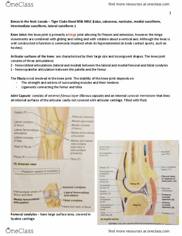
The distal end of the femur ends in two spirally curved femoral condyles (medial and lateral). The femoral condyles articulate with the tibial condyles to form the knee joint. Figure 5. 2 anterior view because you can see the patella fibula is always on the lateral side. Posterior view no patella can see how much higher the femoral cartilage raps around. Is a large sesamoid bone that is formed intratendinously after birth (babies have no knee caps) Posterior surface smooth, articular cartilage, vertical ridges (separates medial and lateral) This triangular bone, located anterior to the femoral condyles, articulates with the patellar surface of the femur. Tibia: the large weight bearing tibia (shin bone) Articulates with the femoral condyles superiorly, the talus inferiorly. Tibial tuberosity site of muscle attachment and the fibula laterally at its proximal and distal ends. The distal end of the tibia is smaller than the proximal end and has facets for articulation with the fibula and talus.