MBIO 1010 Lecture Notes - Lecture 7: Gram Staining, Tetrapeptide, Gram-Positive Bacteria

8 May 2017
School
Department
Course
Professor

Microbiology 1010 – Lecture Notes
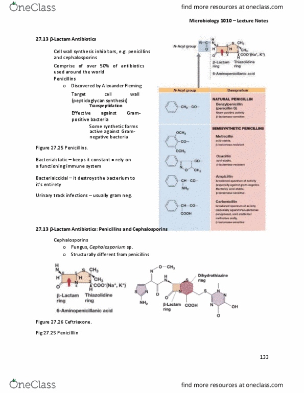
Role of the Cell Wall
Cell wall prevents cell expansion – protects against osmotic lysis
Protects against toxic substances – large hydrophobic molecules
o ex. detergents, antibiotics
Pathogenicity (when have a capsule)
o Helps evade host immune system
Helps bacterium stick to surfaces
Partly responsible for cell shape.
Why do we salt fish? It makes a hypertonic environment – which means the bacteria on the fish
dehydrate and shrink.
Peptidoglycan
Species of Bacteria separated into two groups based on Gram stain
Gram-positives and gram-negatives have different cell wall structure (Figure 2.24)
Gram-negative cell wall
Two layers: LPS and peptidoglycan o
Gram-positive cell wall
One layer: peptidoglycan
Endospores – only from gram positive (but not all gram positive)
All gram neg. have lipid and sugar. Lipid is on the outer membrane and the sugar is then attached
to that. Together it is called LPS (lipopolysaccharide) = endotoxin.
LPS – is ‘o’ linked. It helps us to determine different features of bacteria.
It’s called an exotoxin = secreted to the outside
Endotoxin – is physically attached.
Once LPS is released from the dead cell, it can cause shock from the vassal dilation and can cause the
person to die. Which is why depending on the gram neg. bacteria infection, antibiotics won’t be given.
37

Microbiology 1010 – Lecture Notes
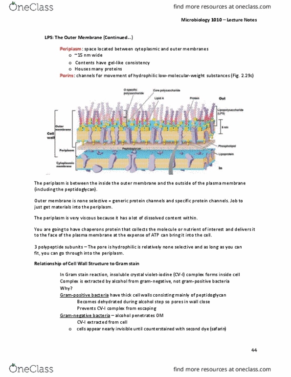
Gram neg:
Periplasmic space = Area of the interior of the outer membrane and the exterior of the
plasma membrane.
In between these two membranes, there is the thin layer of peptidoglycan
38

Microbiology 1010 – Lecture Notes
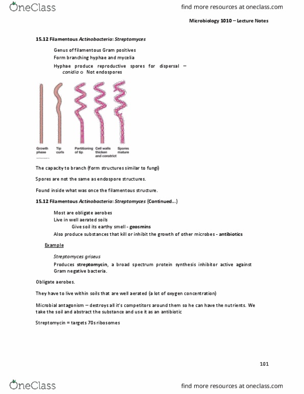
Peptidoglycan (Continued…)
Rigid layer that provides strength to cell wall
Polysaccharide composed of:
o N-acetylglucosamine (NAG) and N-acetylmuramic (NAM) acid
o Amino acids
o Lysine or diaminopimelic acid (DAP)
o Cross-linked differently in gram-negative bacteria and gram-positive bacteria
o Form glycan tetrapeptide
There is many different variations that are found within gram neg and positive bacteria.
NAM and NAG are the disaccharides when coupled together to make majority of the structure.
Creating a lattice.
The D form is unique to this case to the bacteria and few other exceptions.
The 5 glycine there is alternative amino acid threads to bridge the gap between the structure –
this glycine is specific for staphylococcus aureus.
39
Document Summary
Cell wall prevents cell expansion protects against osmotic lysis. Protects against toxic substances large hydrophobic molecules: ex. detergents, antibiotics. Pathogenicity (when have a capsule: helps evade host immune system. It makes a hypertonic environment which means the bacteria on the fish dehydrate and shrink. Species of bacteria separated into two groups based on gram stain. Gram-positives and gram-negatives have different cell wall structure (figure 2. 24) Endospores only from gram positive (but not all gram positive) Lipid is on the outer membrane and the sugar is then attached to that. Together it is called lps (lipopolysaccharide) = endotoxin. It helps us to deter(cid:373)i(cid:374)e differe(cid:374)t features of (cid:271)a(cid:272)teria. It(cid:859)s (cid:272)alled a(cid:374) e(cid:454)oto(cid:454)i(cid:374) = se(cid:272)reted to the outside. Once lps is released from the dead cell, it can cause shock from the vassal dilation and can cause the person to die. Which is why depending on the gram neg. bacteria infection, antibiotics won(cid:859)t (cid:271)e gi(cid:448)e(cid:374).