BIOL110 Lecture Notes - Hox Gene, Aortic Arch, Trochlear Nerve

30 Jan 2013
School
Department
Course
Professor
Document Summary
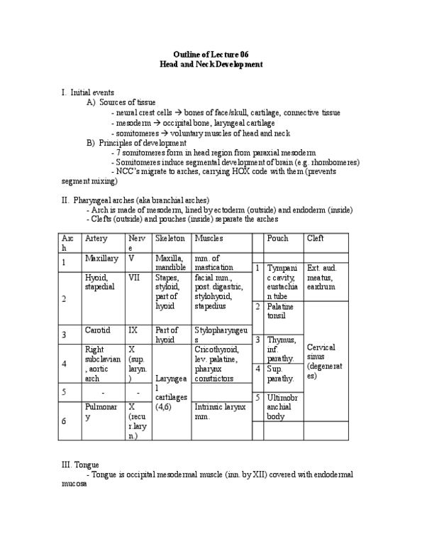
Somites 2-5 (note: raymond has replaced a table image with equivalent text) Summary of info, thus far: each segment of neural tube has unique genetic identity (hox) carried into pharyngeal arches through neural crest cells some cells die to for grooves coordinate brain and facial development. 5th (note: raymond has replaced a table image with equivalent text below) 5 pairs first pouch second pouch third pouch. 1st pharyngeal cleft penetrates underlying mesenchyme and forms eam: bottom of eam forms lateral aspect of tympanic cavity, undergoes active proliferation and overlaps remaining clefts, merges with ectoderm of lower neck remaining cleft have no contact. 2nd cleft with outside: temporarily, clefts form cervical sinus, but disappears. Thyroid gland: arises from foramen cecum, descends along front of pharyngeal gut, remains connected to tongue by narrow canal: thyroglossal duct (obliterated later, descends just caudal to laryngeal cartilages, functions during early fetal period.