CAPS 391 Lecture Notes - Lecture 35: Levator Palpebrae Superioris Muscle, Extraocular Muscles, Ciliary Muscle

18 Nov 2015
School
Course
Professor

Document Summary
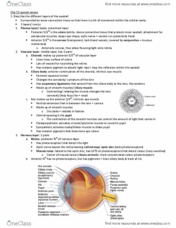
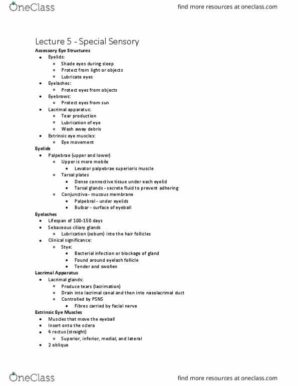
Levator palpebrae superioris muscle: skeletal muscle, attached to the upper eyelid and functions to raise. Innervated by branches from cniii, as well as from the sympathetic nervous system (fibers attached to tarsal plate). If there is a problem with the sympathetic nervous system - - > drooping of eyelids (ptosis: cornea: anterior side of eyeball. Nerves and blood vessels penetrate to gain access to deeper layers. Second layer, deep to sclera (vascular): posterior 5/6 is the choroid (supplies blood for retina). Far vision: ciliary muscles relax (sns), the suspensory ligaments stretch and the lens flattens. Iris is a disc covering the anterior side of lens. Sympathetic innervations dilate pupil, and parasympathetic constrict. Innermost layer (nervous layer): retina is the posterior 3/4. The anterior 1/4 is a pigmented layer. The center of it is a dark area called the fovea centralis, with the highest concentration of photoreceptors (cones). Aqueous humor flows through the pupil from posterior chamber to the anterior chamber.