CAPS 391 Lecture 36: Lecture 36 summary

18 Nov 2015
School
Course
Professor

Document Summary
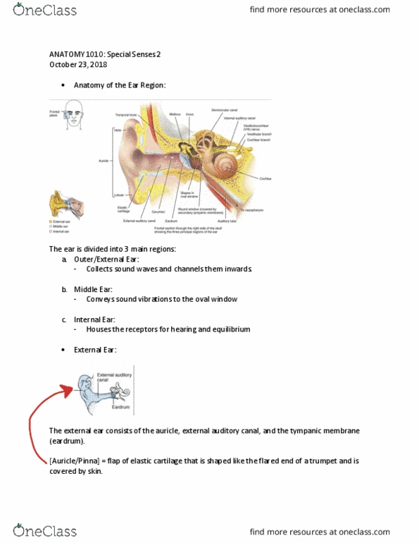
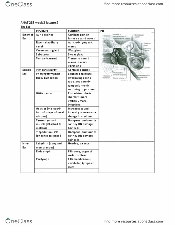
Auricle: formed by elastic cartilage apart from the lobe (loose ct). External acoustic meatus (1 inch): mainly elastic cartilage apart from the medial side (bone). It conducts the sound and produces cerumen to protect the ear. Tympanic membrane (1cm2): limit between external and middle ear. It has a shallow depression at the centre called umbo. Tympanic cavity has 6 surfaces: roof, lat wall, medial wall and floor (the other 2 are not examinable) 3 bones hold at the surfaces by ligaments and articulated by synovial joints: malleus (attached to umbo), incus and stapes. 2 skeletal muscles prevent hypervibration: tensor tympani (attached to malleus, cn v) and stapedius (attached to incus, cn vii) 2 windows continuous with the internal ear: oval (attached to bone to conduct vibration) and round (not attached to bone to stop vibration) Mastoid sinus: extension of the tympanic cavity, continuous with the middle ear (focus of infections)