ANP 1106 Lecture Notes - Lecture 4: External Occipital Protuberance, Condyloid Process, Superior Orbital Fissure

2 Feb 2016
School
Department
Course
Professor
Document Summary
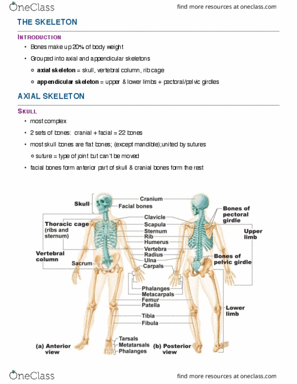
Appendicular skeleton = bones of upper/lower limbs + pelvic girdles [attachment to axial skeletons] Cranium: skull divided into vault: superior part(lateral/posterior) & base: inferior. 3 bony ridges of the base: anterior(high), middle, posterior(low) fossa. Cranium: 8 bones: paired(2) parietal & temporal unpaired(1) frontal, occipital, sphenoid, ethmoid. Curvature of skull gives strong outer surface for protection. Supraorbital margin: end of frontal bone below eye brow. Occipital condyles: on both sides of magnum site for articulation with first cervical vertebra. External occipital protuberance: lines of projection at the back of skull. Temporal bone parts: squamous region: big flattened zygomatic process to cheekbone, tympanic region: surround ear (external acoustic meatus-opening, petrous region: inside temporal bone. Ethmoid bone parts: inside orbits/nasal cavities: cribriform plate: make roof of nasal cavity (olfactory foramina: tiny hole for olfactory nerves, perpendicular plate: points downward -part of nasal septum, crista galli: points upward attachment part of brain. Ethmoid sinuses: on the lateral part of ethmoid bone.