HSS 1100 Lecture Notes - Lecture 3: Bunsen Burner, Agar Plate, Freeze-Drying

30 Apr 2018
School
Department
Course
Professor

Diagnostic Microbiology
• Microorganisms exist as mixture cultures within the environment. To study them you
must isolate the specific organism you want to look at allowing you to view its
characteristics.
• Some things you would need: Bunsen burner, Agar plate
• Inoculation methods: how you will get the bacteria to colonize enough to be seen
o Pour, Spread, Streak
o Most common method is the Streak plate method
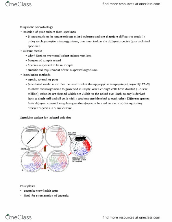
Streaking a plate for isolated colonies
1. Sterilize the inoculation loop with a Bunsen burner
2. Wait for it to cool down and then place the loop in the sample. And then place a small
drop on the agar plate
3. Placed in the incubator to grow and start colonizing. (after 10million cells colonies will
become more visible)
4. The same process is repeated again in a different sample area and placed onto another
area of the agar plate.
Pour Plates
• Pour the sample into the agar allowing the bacteria to grow within the agar
• Used for enumeration (listing) of bacteria
Streak Plates
• A tiny drop of the sample is placed on to the agar
• It is spread around the agar plate and allowed to grow within the incubator
Preservation of Cultures
• Pure cultures are stored within a fridge (frozen at -80C)
• 3 Methods of storage (Short term vs Long term Storage)
o Frozen in Liquid Nitrogen (-195C)
o Frozen in special freezer (-70C to -120C)
o Lyophilization (freeze-drying) : dehydration and vacuum sealing
Why would we want to keep a copy of a bacteria that was isolated from a patient?
Can be used to monitor outbreaks in future years
Identification
• Ways you can identify pure cultures after isolating them…
o Colony morphology
o Cell morphology

• Using a microscope on High resolving power you have the ability to look at the two
microorganisms that are beside one another and distinguish them as being separate and
being distinct entities
Colonial Morphology
• By looking at the microorganism without using a microscope what do they look like
• Using the criteria: FORM, ELEVATION, MARGIN
Cell Morphology
Staining Techniques
• 3 steps for staining
1. Place the sample on a glass slide
2. It is going to be fixed on the glass slide by passing heat from the Bunsen burner
3. Stain the sample with desired dye
• Simple vs. Differential staining
o Simple stain
▪ Staining of microorganisms using a single dye. Just trying to figure out if
there are organisms present within a sample and look at their general
shape, size, number. Doesn’t give a lot of information.
o Differential stain
▪ Using two or more dyes to see the differences in the organism’s present.
▪ Ex. Acid fast, Gram stain
Gram Stain (Hans Christian Gram)
1. Flood the slide with Crystal violet
2. Flood with Iodine treatment
3. Decolorization with 95% ethanol
a. Very important to only leave on for 20sec bcs that’s all it takes to remove the
crystal violet from a Gram negative cell wall
4. Flood with safranin (pink color)
Gram + → Stain Purple Gram - → Stain Pink
Cell Wall is Key!
• Cell wall is a rigid structure that gives the bacteria its shape
o Shape is related to peptidoglycan layer
• Needed for cell growth and division
• Gram – is thinner than Gram +
• All Staining solution bind to the peptidoglycan of the cell wall
• Gram +
o Main component is peptidoglycan which is very thick


11
HSS 1100 Full Course Notes
Verified Note
11 documents
Document Summary
Diagnostic microbiology: microorganisms exist as mixture cultures within the environment. To study them you must isolate the specific organism you want to look at allowing you to view its characteristics: some things you would need: bunsen burner, agar plate. Inoculation methods: how you will get the bacteria to colonize enough to be seen: pour, spread, streak, most common method is the streak plate method. Streaking a plate for isolated colonies: sterilize the inoculation loop with a bunsen burner, wait for it to cool down and then place the loop in the sample. Pour plates: pour the sample into the agar allowing the bacteria to grow within the agar, used for enumeration (listing) of bacteria. Streak plates: a tiny drop of the sample is placed on to the agar. It is spread around the agar plate and allowed to grow within the incubator. Can be used to monitor outbreaks in future years.