PSY 3301 Lecture Notes - Lecture 2: Inferior Frontal Gyrus, West Nile Fever, Lateral Sulcus

25 Jan 2017
School
Department
Course
Professor
Document Summary
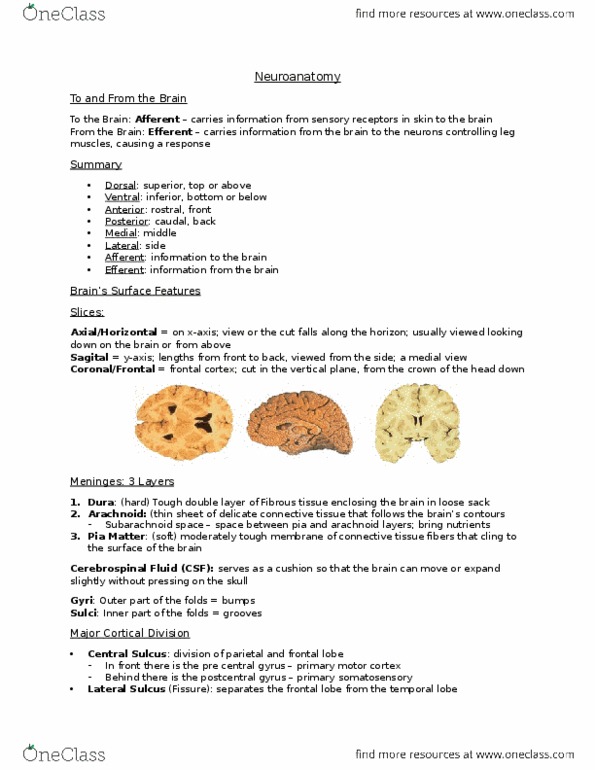
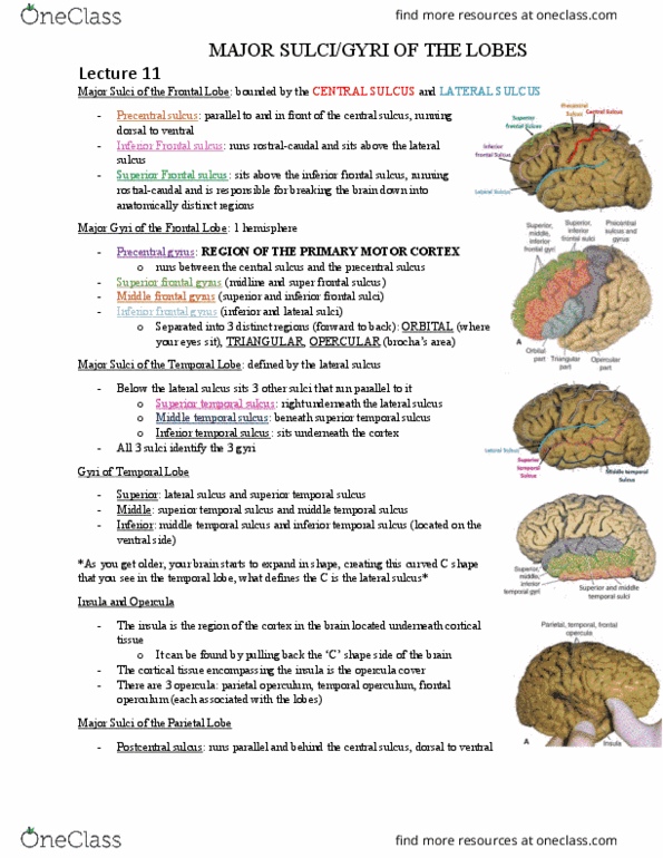
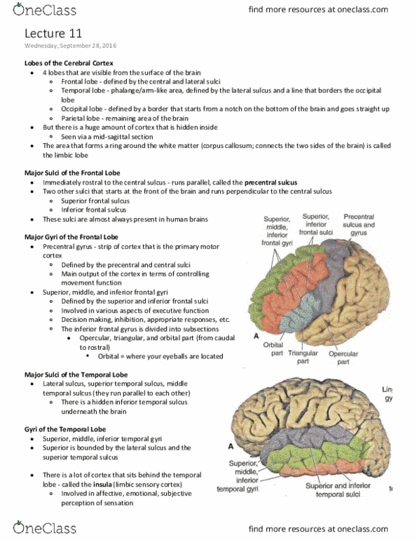
Bigger gap between the hemispheres at the back of the brain, less space between the back of the brain and the skull. Dorsal, lateral, medial, axial (horizontal), sagittal, coronal (frontal section), Must draw a picture for the first examination. Encephalitis is an infection in the actual brain. Only sulcus that goes all the way lateral from the hemispheric space. Post central gyrus: closer to the back of the brain (somatosensory cortex) and; precentral gyrus: closer to the front (primary motor cortex resides) postcentral and precentral sulcus lateral sulcus (temporal to frontal lobes) Superior, inferior, middle frontal and inferior frontal gyrus. Anterior: feeds the top and ront of brain. Posterior: ventral and posterior part of the brain. If you had a stroke, dependent on where is occurred, you will have different symptoms. Gyri: nuclei and dendrites (grey) axons (white matters) Midline that connects to the 4th ventrical, being most ventral. Spi(cid:374)al ta(cid:271)s tells the (cid:374)utrie(cid:374)ts a(cid:374)d (cid:373)eta(cid:271)olites i(cid:374) o(cid:374)e"s (cid:374)ervous syste(cid:373)