BIOD27H3 Lecture : Textbook notes-Chapter 9-The Central Nervous System

11 Aug 2010
School
Department
Course
Professor


MAMMILIAN PHSYSIOLOGY
Chapter 9 ± the central nervous system
Affective Behaviours: Behaviours that have to do with feelings and emotions
Cognitive Behaviours: Behaviours related to thinking
The CNS consists of the brain and the spinal cord
Early Development
In the very early embryo, the cells that become the CNS lay flattened on a region called the neural plate.
At about 20 days old, the neural plate starts migrating towards the midline to form a hollow neural tube.
By 23 days the neural tube is almost complete.
The neural tube remains hollow and eventually will become the CNS. The cells in the inside of the tube
either become ependyma (connective tissue that separates fluids) or undifferentiated stem cells. The
cells on the outside become the neurons and the glial cells. The neural crest cells become the neurons
of the PNS.
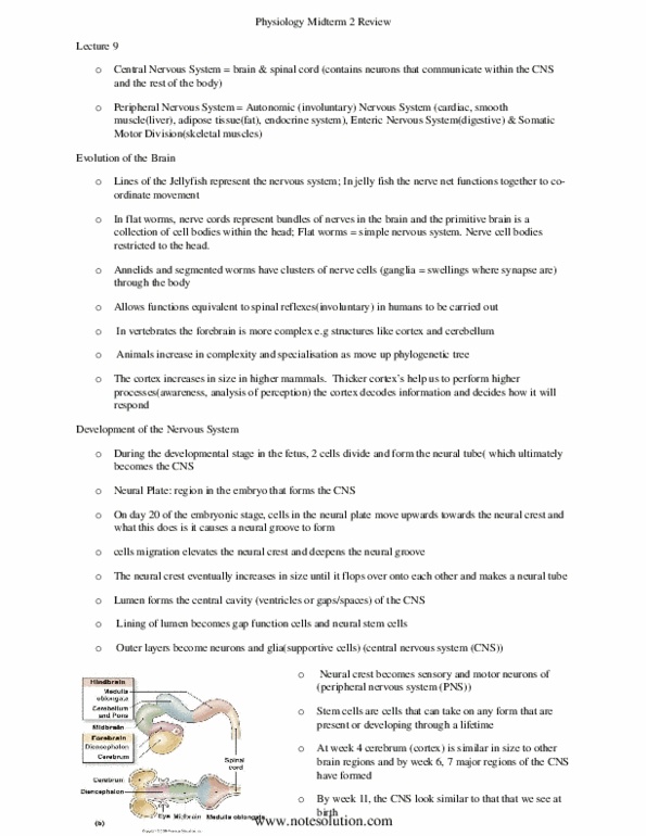
By 4 weeks, 3 distinct divisions of the CNS are obvious: the forebrain, the midbrain, and the hindbrain.
At this point, the forebrain is no bigger than the other parts
By week 6, growth of the forebrain outpaces the other regions and the embryo has developed all 7
regions of the CNS present at birth: the cerebrum, the diencephalon, the midbrain, the cerebellum, the
Pons, the medulla oblongata, and the spinal cord
Also by week 6, ventricles form and the neural tube becomes the central canal of the CNS
By week 11, the cerebrum is much more developed than the other regions and is the most obvious
structure when the infant is born
Grey & White Matter
The tissue of the CNS are divided into 2 groups: white matter and grey matter
Grey Matter: Grey matter consists of unmyelinated nerve cell bodies, dendrites, and axons
terminals. They usually form layers over certain parts of the brain or form
clusters of neurons with the same function (called nuclei)
www.notesolution.com

White Matter: White matter is made up mostly of myelinated axons and contains very few cell
bodies. The white colour comes from the myelin (bundles of axons that connect
to different regions of the CNS are known at tracts. Tracts = nerves in the PNS)
Bone and Connective Tissue Support of the CNS
The brain is encased in a bony skull/cranium and the spinal cord is protected and runs through the
vertebral column (many vertebrae separated by disks of connective tissue)
There are 3 layers of connective tissue in between the spinal cord and the vertebral column: the Dura
Mater, the Arachnoid Membrane, and the Pia Mater (together are called the meninges)
From the bone to the spinal cord, the order is:
1. The Dura Mater: a thick durable layer that is associated with veins that drain blood from
the brain via vessels called sinuses
2. Arachnoid Membrane: A thin cobweb like membrane found between the Dura mater and the
Pia mater
3. Pia Mater: The layer directly on top of the spinal cord, it is associated with the
supply of blood to the brain via different arteries
~Extracellular fluid helps cushion the delicate neural tissue.
Interstitial fluid is found inside the Pia Mater; cerebrospinal fluid is found between the Pia Mater and
the Arachnoid Membrane
Cerebrospinal fluid is a salty solution that is secreted by the choroid plexus (found on the walls of the
ventricles). From there it is pumped into the subarachnoid space (between the Arachnoid Membrane
and the Pia Mater) where it flows around the neural network until it is reabsorbed by villa
It provides physical and chemical protection of the CNS
Physically, it supports the CNS by reducing the weight of the brain bv suspending it in fluid (the
buoyancy of the fluid reduces the weight of the brain by almost a 30 fold). This results in less pressure
on nerves and vesicles. It also provides padding protection reducing damage to the CNS after a blow to
the head or back
Chemically, it supports the CNS by providing a clean environment for the CNS. The choroid plexus is very
selective of the ions and nutrients it passes from the blood. Therefore the cerebrospinal fluid contains
lower levels of K, Ca, HCO3, glucose, and about the same level of Na as the blood.
CSF also exchanges solutes with interstitial fluid of the CNS and provides routs by which waste can be
disposed of
www.notesolution.com

Blood-Brain Barrier
Most of the capillaries in the CNS form the blood brain barrier which protects the brain and spinal cord
from fluctuations in hormones, ions, neuroactive substances, toxins, and pathogens.
The capillaries in the brain are much less permeable than other capillaries in the body because they
form tight junctions (with the help of astrocytes), unlike capillaries of the body which use loose, leaky
junctions. Tight junctions do not let anything pass
The capillaries are also very highly selective, only allowing substances to enter via transport proteins
Some areas of the brain do not have a functioning blood-brain barrier. Usually these areas need to
interact with blood to do their job so they do not need a barrier
I.e. the posterior pituitary gland does not have a function blood-brain barrier because its job is
to release hormones via the blood therefore it needs to come in contact with capillaries to
transfer the hormones to the blood
Another example is the vomiting center in the medulla oblongata. It does not have a functioning
blood brain barrier because it needs to constantly monitor blood to see if there is are toxins or
foreign substances.
In terms of nutrients, glucose is really the only nutrient for neurons of the CNS. About 15% of the blood
pumped by the heart and about 50% of the oxygen in the body goes to the brain
*The Spinal Cord (***see fig. 9-7 on page 302 for this part***)
The spinal cord is the main pathway for communication between the brain and the skin, joints and
muscles of the body
Also it is responsible for movement and sensation; if the spinal cord is severed it can lead to a loss of
sensation and paralysis
The spinal cord is divided into 4 sections: cervical, thoracic, lumbar, and sacral. Each region is subdivided
into segments which have their own bilateral pair of spinal nerves.
Right before the spinal nerves join the spinal cordUZ]À]]v}Á}Z}}[~Zdorsal and ventral
roots)
The dorsal root of each spinal nerve is specialized to carry incoming sensory information (from
the PNS to the CNS). Dorsal roots contain large swellings of bundled sensory nerves just before
they join the spinal cord (called dorsal root ganglion)
The ventral root carries information from the CNS to the PNS (efferent motor and autonomic
neurons)
www.notesolution.com
Document Summary
Behaviours that have to do with feelings and emotions. The cns consists of the brain and the spinal cord. In the very early embryo, the cells that become the cns lay flattened on a region called the neural plate. At about 20 days old, the neural plate starts migrating towards the midline to form a hollow neural tube. By 23 days the neural tube is almost complete. The neural tube remains hollow and eventually will become the cns. The cells in the inside of the tube either become ependyma (connective tissue that separates fluids) or undifferentiated stem cells. The cells on the outside become the neurons and the glial cells. The neural crest cells become the neurons of the pns. By 4 weeks, 3 distinct divisions of the cns are obvious: the forebrain, the midbrain, and the hindbrain. At this point, the forebrain is no bigger than the other parts.