BIOC19H3 Lecture Notes - Lecture 7: Paraxial Mesoderm, Myocyte, Neural Tube


Document Summary
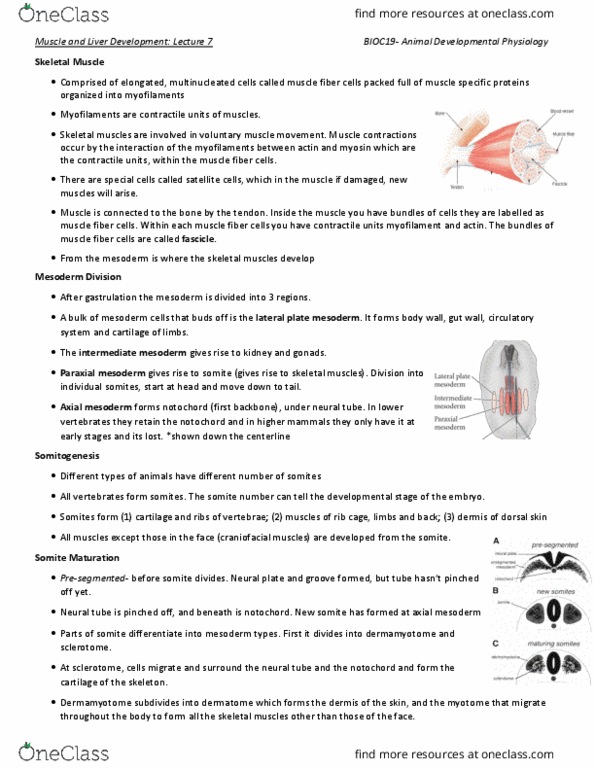
Slide 2: skeletal muscles a. k. a voluntary muscles, myofilaments are composed of muscle specific proteins: actin and myosin. Slide 3: pink diagram shows the development of the neural tube. The ectoderm folds-in, forms the neural groove, and then the neural tube pinches out. This diagram shows the cross-section of the neural tube: black diagram shows the top view of the neural tube. The early neural groove is not zipped and the neural tube on its right is zipped up or closing neural tube in its late phase. Its starts to zip at the brain and then to the posterior end of the embryo. Neural tube zippers up before the development of muscles: the diagram on the lower left also shows a cross-section of a neural tube. Right under the neural tube is notochord and the red part is mesoderm, which eventually develops into different regions. The red mesoderm cells proliferate and surround the embryo.