PSYB65H3 Lecture Notes - Headache, Gyruss, Subarachnoid Space

Document Summary
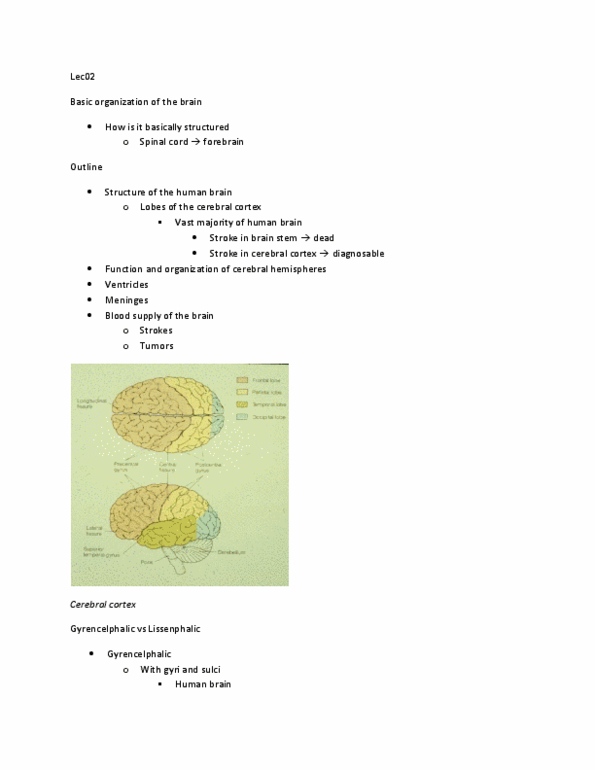
Lecture 2 function of the cerebral hemispheres, brain vascular supply, cerebral ventricles, cerebral coverings (chapters 2, 14) Organization and structure of the human brain, especially the cortex. Function of the lobes and the functions within each lobe. Outer structure surrounding the brain (meninges), which are the coverings of the brain. Inner structure of the brain such as the ventricles. As you move forward in the brain, the cortex is the newest evolutionarily. The cortex is not smooth for humans. There is some evolutionary pressure that wants humans to be smart. Sulcus/sulci: the valleys in the brain. If there are huge (major one), it"s called fissure. Gyrus/gyri: the individual mountains in the brain. If you look at the top of the brain, the 2 sides of the brain are not connected from the top view, but they actually are connected. The brain is divided into two hemispheres (right and left), but they are not indentical.