ANA300Y1 Lecture 13: Lecture 13- Meninges and the Cerebral Hemispheres.pdf

Document Summary
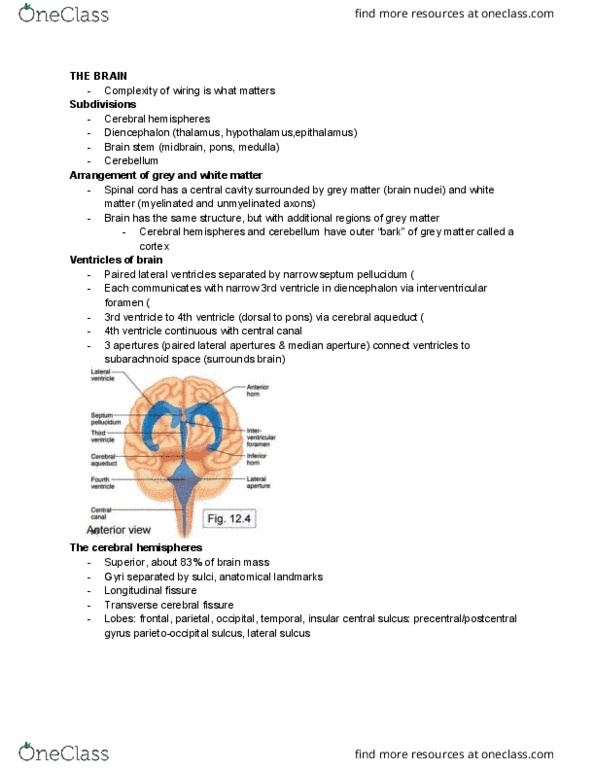
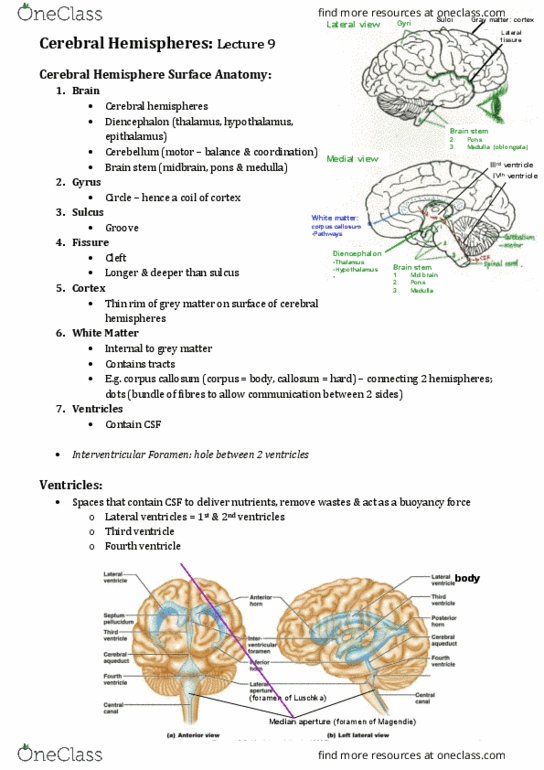
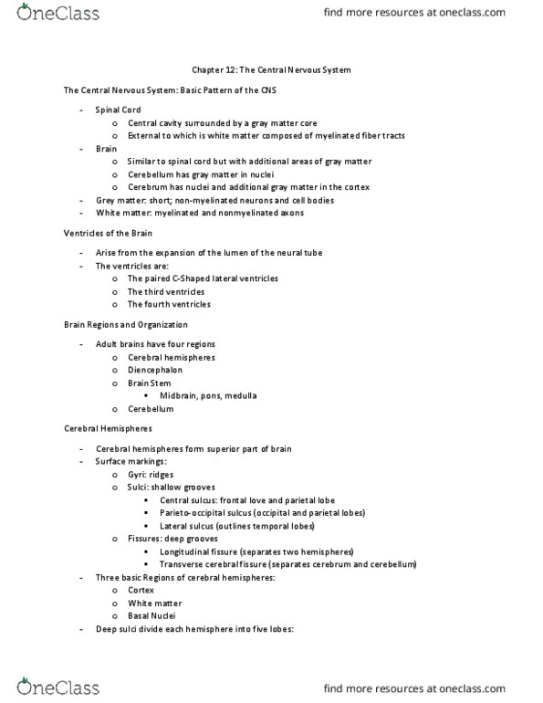
Lecture 13: meninges and the cerebral hemispheres. Lobes of the cerebral hemispheres preoccipital notch lateral fissure. Dural venous sinuses and cerebral venous. Meets up with tentorium cerebelli at the back forms a roof over the cerebellum. 3rd ventricle: empties via cerebral aqueduct: narrow communication that passes through the midbrain, opens up to the 4th ventricle. Continuous with the 3rd ventricle. Choroid plexus in the lateral ventricles. Csf percolates around the surface of the cerebrum, cerebellum, down in the subarachnoid space spinal cord. Ultimately returned to venous blood via. Some csf can be absorbed into lymphatics surrounding cranial and spinal nerves by flowing through the sheaths surrounding the nerves. Brain doesn"t have lymphatics, but there are lymphatics in the connective tissues surrounding the nerves. Right side: ventricles are enlarged occurs because flow of csf is blocked (ex. cerebral aqueduct is clogged ) compresses surrounding neuron tissue. Function in programming of, preparation for, movement and control of postural adjustments.