BIO130H1 Lecture Notes - Lecture 19: Immunofluorescence, Electron Microscope, Refraction


31
BIO130H1 Full Course Notes
Verified Note
31 documents
Document Summary
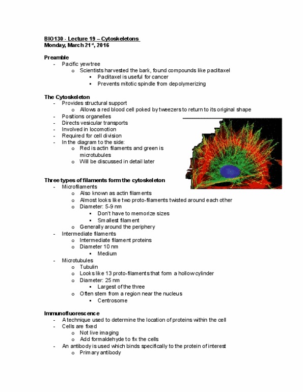
Microfilaments kind of looks like 2 protofilaments twisted around eachother. Microtubules look like 13 protofilaments that form a hollow structure. Actin filaments are very often near the periphery. Microtubules stem from an area near the nucleus, known as a center called the centrosome. The secondary antibody binds to the first one, and it has a fluorescent molecule that lights up when viewed under a fluorescent miscoscope. You could also use primary antibodies with the fluorescent molecule already on it, but this way is cheaper. Refraction is the bending of light or waves as it passes through obstacles. With an electron microscope you can look at a more detailed structure because you can turn the image. The alpha and beta form heterodimers and they both bind to gtp. Then the tubulin heterodimers will start binding to eachother. What is formed going up and down is called a protofilament, and 13 protofilaments form a microtubule.