CSB351Y1 Lecture Notes - Lecture 10: Rna Virus, Pancreatic Ribonuclease, Ribonuclease

26 May 2018
School
Department
Course
Professor

Lecture 10: RNA Virus Replication
• Outside a living cell, viruses are usually inert
• They depend entirely upon the host cell for the supply of precursors and for use of the cell machinery (e.g
ribosomes, tRNAs, etc)
• Oe iside the host ell, iuses ake a kid of oup detat to take oe ost of the ells ahiey
• Inhibition of some cellular metabolic pathways is a common strategy
• Nucleotides, AAs, ATP and free ribosomes become readily available
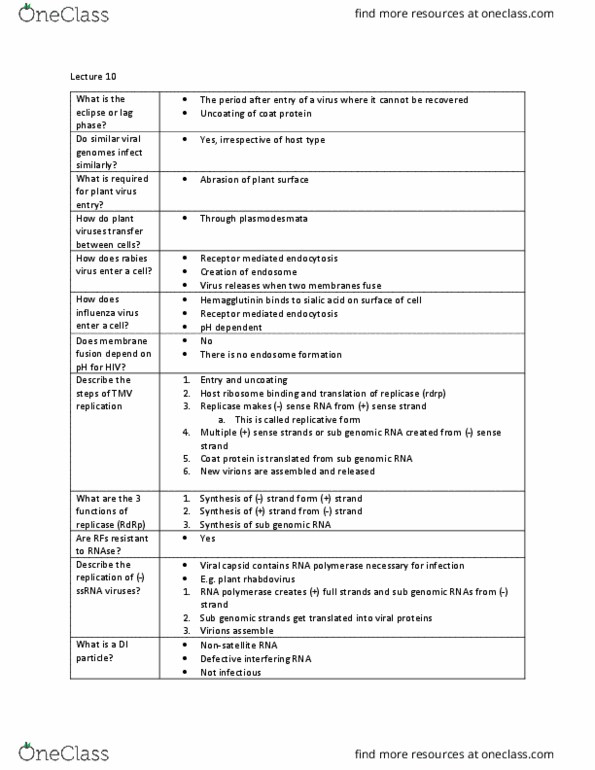
Stages of virual infection
1. Absorption and entry
2. Elipse of lag epliatio
3. Maturation and assembly
4. Movement, storage and release (exit) in some cases
Virus Replication Cycle
• Analysis of viral macromolecules reveals the detailed pathways of virus
replication
1. Eclipse (lag) Phase
• Following entry of the viral genome into a
host cell, there is a period in which
infectious virus cannot be recovered from
the cell – eclipse or lag period
- Most of the virus components are
synthesized de novo (from nothing)
• Viruses with similar types of genomes
usually replicate in similar ways,
irrespective of which type of organisms
they infect
- Replication of SS (+) RNA viruses are
similar in plants, animals and bacteria
Absorption and Entry of Plant Viruses
• Plant cells have thick cell walls – it is necessary to wound the cell wall to allow infection to take place
- Epidermal hairs are broken by abrasion or wound
• Vectors (e.g. insects) act in the same way – virions enter the cell within membrane bound vesicles (pinocytosis)
- Plant cell wall acts as very effective barrier to virus entry into cells
• Plant RNA virus – at the same time or soon after entry, the virus is disassembled (uncoating)
- Virus particles disappear from view from pinocytic vesicles soon after entry into the cell – eclipse stage
• Uncoating may play a part in the control of host range
- e.g. BMV-RNA coated with TMV-protein failed to uncoat when infected its host cells – no infectivity, no
release of RNA
Plasmodesmata create gaps that connect plant cells
find more resources at oneclass.com
find more resources at oneclass.com

Some experimental evidence for uncoating of TMV
• Viruses on inoculation become sensitive to UV radiation
- susceptibility decreases with time (RNA when uncoated
becomes sensitive to UV)
• Newly formed TMV particles, after inoculation, are detectable
24 hours sooner when RNA is used as inoculum
- Time difference corresponds to the time taken for
uncoating
• RNase sensitive RNA appears after approx.. 15min of inoculation with TMV particles
(use of 32 p)
• 15-20% of the RNA is released from the virus within 7 min. after inoculation
• Symptoms appear on plats several hours earlier if naked RNA is used for inoculation
• RNase A prevents infection of TMV if it is rubbed on leaves just after inoculation – no effect after few hours
• Photo-reactivation of RNA is possible if the inactivation with UV is carried out directly after inoculation – no
reaction after 15mins
Absorption and Entry of Animal Viruses
• In human and animal viruses, viruses recognize specific cells
thorugh the recognition of molecules found on their surface –
receptors (CD = cluster of differentiation)
Virus entry and receptors
• Variety of cell surface proteins can serve as specific virus
receptors or attachment factors
• PVR = Poliovirus Receptors (CD155)
• CAR = Coxsackie virus and Adenovirus Receptor
• LDL = Low Density Lipoprotein
• Receptor-mediated endocytosis
- Virus receptor binds to virus, endocytosis,
endosome, fusion, release of viral RNA
- i.e. Rabies cycle
- can also be pH dependent
Penetration through cellular membranes
• Fusion proteins undergo major conformational changes that lead to fusion
- Activated by low pH or receptor binding
Entry of Influenza Virus
find more resources at oneclass.com
find more resources at oneclass.com