PSL300H1 Lecture Notes - Lecture 3: Optic Chiasm, Central Retinal Artery, Ciliary Muscle

Document Summary
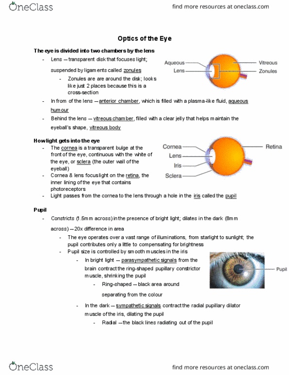
The eye is divided into 2 chambers by the lens. The lens is a transparent disk that focuses light. In front of the lens is the anterior chamber, filled with aqueous humor, Behind is the vitreous chamber, filled with the vitreous body, plasma-like fluid. maintain the eyeball"s shape. a a clear jelly that helps. Light enters the eye through the cornea. Transparent bulge at the front of the eye, continuous with sclera the outer wall of the eyeball. Cornea and lens focus light on the retina, the inner lining of the eye that contains the. Light passes from the cornea to the lens through a hole in the iris called the pupil. photoreceptors. In bright light: constrict (shrink) to 1. 5 mm across , In the dark: dilate (enlarge) to 8 mm , ~20 times bigger in area. Reducing the amount of light reaching the lens.