PSL301H1 Lecture Notes - Lecture 15: Thoracic Cavity, Intrapleural Pressure, Thoracic Wall


41
PSL301H1 Full Course Notes
Verified Note
41 documents
Document Summary
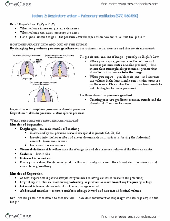
Inspiration: brainstem signals phrenic nerve, inspiratory muscles contract thoracic volume increases, lung expands pulls air inwards. Expiration: lungs at peak volume activation of stretch receptors, diaphragm stops pulling downwards diaphragm relaxes, chest contracts air goes out. Pneumothorax: ribcage & thoracic cavity expand, sternocleidomastoids & scalenes contract, diaphragm muscle contracts & moves down, thoracic cavity pressure decreases & air fills, pressure inside lungs decrease air moves in lungs expand. Liquids = non-expandable: lungs follow volume changes of the throat, intrapleural pressure create a negative pressure (-3 mm hg, pleura = suction cup. Pleural membranes adhere to lungs & thoracic wall: via connective tissue. Definition: air in the pleural cavity disrupts negative pressure bonding lung to chest wall. The chest wall expands outwards: no longer bound by pressure to the lungs. Wet dressing on the wound (act as a one-way valve) Apply pressure at mouth to inflate lungs. Severe cases: surgery to remove & repair damaged pleura.