Anatomy and Cell Biology 2221 Lecture Notes - Lecture 5: Long Thoracic Nerve, Serratus Anterior Muscle, Axillary Artery

27 Sep 2018
School
Department
Professor
Document Summary
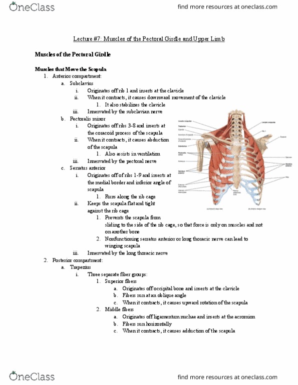
Attachments: clavicle, sternum, and upper costal cartilagesto the bicipital groove. Attachments: junction of 1st rib & its costal cartilage to the groove on middle 1/3 of clavicle. Movement: protraction, upward rotation, holds scapula onto the thoracic wall. Winged scapula: when there is damage to serratus anterior or lo(cid:374)g thora(cid:272)i(cid:272) (cid:374)er(cid:448)e, the s(cid:272)apula (cid:272)a(cid:374) (cid:862)(cid:449)i(cid:374)g(cid:863) out fro(cid:373) ri(cid:271) (cid:272)age as it (cid:272)a(cid:374)"t (cid:271)e held o(cid:374)to it. Attachments: med. ant. border of the scapula onto ribs 1-8. This is a pyramid shape, fat filled space located b/w the lateral wall of the upper thorax and your upper arm. It acts as a passageway for blood vessels and nerves in the axillary sheath: Pec : axillary vein, axillary artery, brachial plexus cords. This provides the upper limb and part of thoracic wall with blood. This is a complicated artery as it has many parts: 1st part = single branch, the superior thoracic artery.