Anatomy and Cell Biology 3309 Lecture Notes - Lecture 34: Outer Plexiform Layer, Retinal Pigment Epithelium, Inner Plexiform Layer

22 May 2018
School
Department
Professor

Histology 3309
Eye II
Objectives
1. Understand the organization of the retina
2. Compare and contrast rod and cone photoreceptor cells
3. Understand the overview of the retinal circuitry
4. Understand what fovea, macula and optic disk are
The Eye
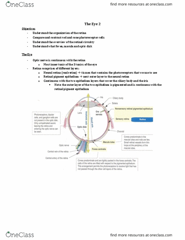
- The retina is made of…
o the neural retina (tissue that contains the photoreceptors that we use to see with) and
o the retinal pigment epithelium
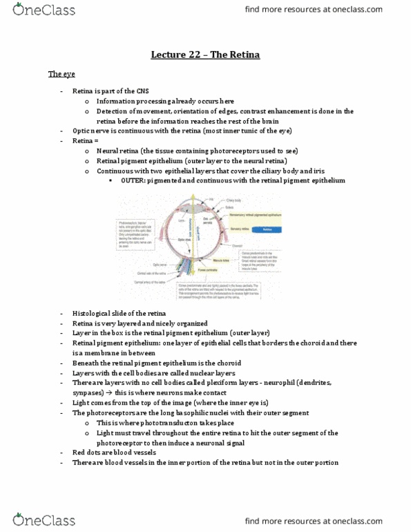
- this is the retina
- the layer at the bottom is the retinal pigment epithelium which bords the choroid (contains all
the blood vessels) and the sclera would be even more outer to that
- layers with cell bodies = nuclear layers
- layers without cell bodies (containing neurophil) = plexiform layers
- white space at top is the center of the eye
- red dots in this image = blood vessels
o these are only present in the inner portion of the retina
o NOT PRESENT IN THE OUTER PORTION OF THE RETINA
find more resources at oneclass.com
find more resources at oneclass.com

The Retina
- Layers listed from most outer to inner:
o Retinal pigment epithelial cells
o Photoreceptors
o Outer plexiform layer (synapse of the photoreceptors to the next cells)
o Outer nuclear layer (bodies of photoreceptors)
o Inner nuclear layer (different cells - horizontal cells, bipolar cells, amacrine cells etc)
o Inner plexiform layer (no cell bodies – just contacts)
o Ganglion cell layer (retinal ganglion cells lie here)
▪ These are the only cells that fire action potentials
▪ All the other cells are neurons but they don’t fire APs (they only have graded
potentials)
o Nerve fiber layers (axons of ganglion cells)
The Retinal Pigment Epithelium (RPE)
- Most outer layer
- Border to the choroid
- Functions:
o Contain melanocytes
▪ Meaning that they are pigmented and absorb light
▪ All the light that has not been absorbed is absorbed by these cells bc otherwise,
it would enter the brain/skull
▪ Note: the choroid also absorb some of the excess light
▪ Note: in some animals, the RPE cells reflect the light instead of absorbing them,
allowing them to see at night
o Transport all kinds of nutrients
▪ Photoreceptors cells are neurons so they the RPE cells have to maintain
homeostasis in terms of ion concentrations
o Glial cell functions
find more resources at oneclass.com
find more resources at oneclass.com

▪ Provide nutrients to the photoreceptor cells
o Visual cycle
▪ There is a photosensitive pigment in the outer photoreceptive segment
▪ This photosensitive pigment needs to be constantly recycled bc when a photon
hits it, it undergoes a conformation change which starts a neuron signal
▪ This recycling is done by the RPE cells
o Phagocytosis
▪ Debris from retina
▪ There are some photoreceptors that constantly shed their tips
▪ This forms cell debris that needs to be removed
o Secretion:
▪ Provide nutrients, growth factors etc
- This is EM of RPE cells
o Dark dots = pigmented inclusions
o They have fingerlike protrustions to increase SA
The RPE
- Synthesis of melanin and melanosome formation
- Nutrient transport to outer layers of the sensory retina
- Potassium homeostasis
- Generation of 11-cis retinal
- Phagocytosis of shed disks
- Synthesis of the Bruch’s membrane
o Bruchs membrane plays an important
role in degeneration of the eye
- Blood retina barrier
o RPE cells are attached to each other
via tight junctions
o Nothing can go into the retina that is
from the blood vessels (unless its
lipophilic and can go through the
membrane)
find more resources at oneclass.com
find more resources at oneclass.com
Document Summary
Objectives: understand the organization of the retina, compare and contrast rod and cone photoreceptor cells, understand the overview of the retinal circuitry, understand what fovea, macula and optic disk are. Layers listed from most outer to inner: retinal pigment epithelial cells, photoreceptors, outer plexiform layer (synapse of the photoreceptors to the next cells, outer nuclear layer (bodies of photoreceptors, ganglion cell layer (retinal ganglion cells lie here) Inner nuclear layer (different cells - horizontal cells, bipolar cells, amacrine cells etc) This is em of rpe cells: dark dots = pigmented inclusions, they have fingerlike protrustions to increase sa. Nutrient transport to outer layers of the sensory retina. Synthesis of the bruch"s membrane: bruchs membrane plays an important role in degeneration of the eye. Blood retina barrier: rpe cells are attached to each other via tight junctions, nothing can go into the retina that is from the blood vessels (unless its lipophilic and can go through the membrane)