Anatomy and Cell Biology 3319 Lecture Notes - Lecture 10: Dorsal Root Ganglion, Posterior Grey Column, Conus Medullaris

12 Oct 2016
School
Department
Professor
Document Summary
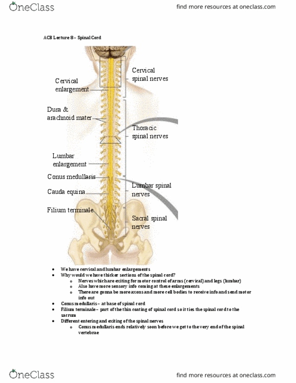
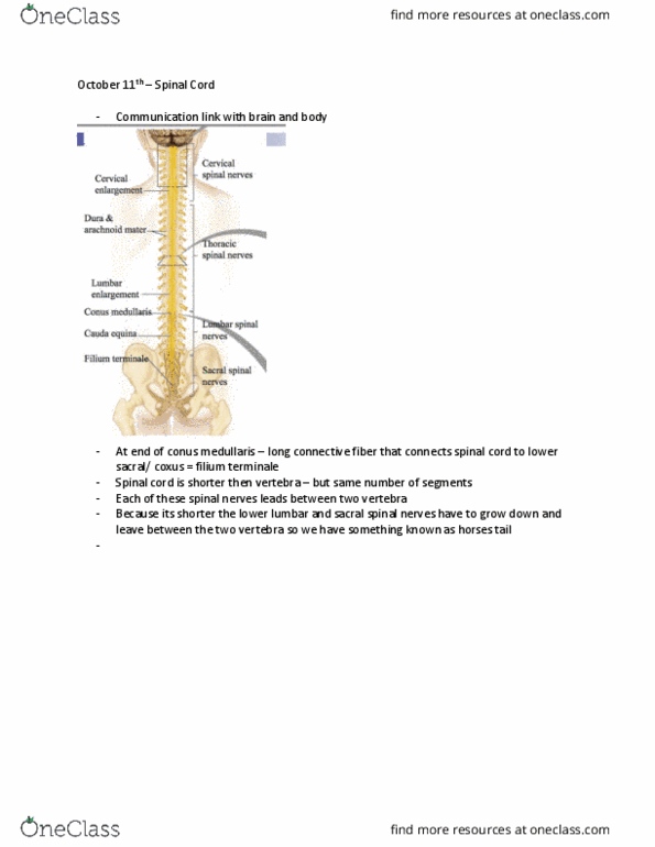
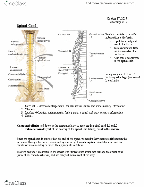
Communication link (how the body communicates info to the brain and how the brain communicates info to the rest of the body) Horseback riding accident severed his spinal cord and is now a paraplegic. After the spinal cord is severed, all of the inputs and outputs below the transection are lost. Two enlargements b/c there extra input and output to the arms and legs. At the end of the conus medullaris there is a connective fiber (filium terminale) that connects the spinal cord to the lower sacral coaxes. The spinal cord is shorter than the vertebra but it has the same number of segments. Each spinal nerve leaves between two vertebra. Because its shorter the lower lumbar and sacral spinal nerves grow down and exit between the appropriate two vertebra resulting in the cauda equina. Image shows the relationship between the spinal cord and the vertebra. Dorsal surface has a gracile and cuneate fasciculus.