Anatomy and Cell Biology 3319 Lecture Notes - Lecture 21: Erector Spinae Muscles, Rib Cage, Internal Intercostal Muscles

1 May 2018
School
Department
Professor

Lecture 021: Muscles of the back, thorax, and neck
Objectives:
● Describe the anatomy of the bony rib cage
● Understand the arrangements and actions of the intercostal muscles and diaphragms
● Describe the intrinsic muscles of the back, including innervation and actions:
○ Deep: transversospinalis
■ interspinales, intertransversarii, rotatores, multifidus, semispinalis
○ Intermediate: erector spinae muscles
■ Spinalis, longissimus, iliocostalis
○ Superficial: splenius
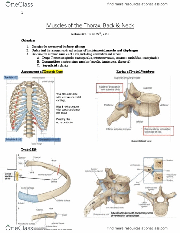
Arrangement of the Thoracic Cage
● Rib cage protect vital thoracic organs (lungs and heart)
● 12 ribs in total:
○ True ribs (1-7):
■ Start at the vertebral column and articulate with sternum via costal
cartilage
○ False Ribs (8-10):
■ articulate with costal cartilage (attach the bone of the ribs to the
sternum) of ribs above
■ Do not directly attach to the sternum
○ Floating ribs (11-12):
■ no articulation with sternum at all
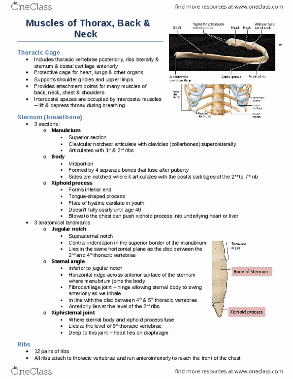
○ A typical rib:
■ Head:
● Attaches to the vertebral body
● Arrow shaped (angular) head fits in between 2 vertebrae (held
together by ligament and wrapped in a joint capsule)
○ 2 articular surfaces
■ Neck:
● Tubercule comes of the neck
○ Points posteriorly
○ Articulates with the transverse process of the inferior
vertebrae
■ Ribs are named for the vertebrae they articulate
with on the transverse process (i.e. the one
below)
■ Body:
● Cross-section: big at superior surface and tapers towards the
inferior surface
● Has a costal groove that runs along the internal surface
■ Ribs ends in costal cartilage
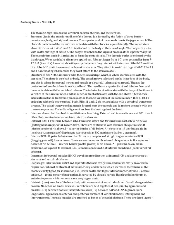
● Sternum
find more resources at oneclass.com
find more resources at oneclass.com

○ A fusion of 3 separate bones:
■ Menubrium
● Has the jugular notch
○ Lateral to this is the clavicle
● Has articular surfaces for the clavicle
○ First rib joins right underneath the clavicle
● Sternal angle
○ Projects up, located beneath the clavicle
○ Important landmark:
■ 2nd rib joins the sternum at this point
○
■ Sternum body
■ Xiphoid process
● Tip of the xiphoid process
○ Marks the end of the rib cage
○ Apex of the abdomen
● Intercostal muscles
○ Helps us breath
○ External intercostal muscle
■ Stretches from the inferior border of the superior rib to the superior border
of the inferior rib
● Fibers goes from an anterior and inferior direction
■ Muscle acts to elevate the rib cage
● Pulls the lower rib up to meet the upper rib
○ Internal intercostal muscle and innermost intercostal muscle
■ The innermost intercostal muscle is a very thin layer
■ Hard to distinguish the two layers in a dissection
■ Both run from the superior border of the inferior rib inferior border of the
superior rib
● Fibers goes from a medial and superior direction
■ Muscle act to depress the rib cage
● Pulls the upper rib to meet the lower rib
○ Innervation and blood supply
■ Neurovascular (nerve, artery, and vein) bundle follows the costal
groove around the rib cage and branch off to the surface
● Costal groove provide some protection
■ Spinal nerve emerges from between the vertebrae
● The anterior/ventral rami tracks all the way to the anterior
section of the body
○ Supplies motor and sensory innervation to the intercostal
muscle
● This gives the striped (dermatome) appearance
■ An artery branches off the aorta at each level of the rib (main)
● Also have collateral circulation from other blood vessels
find more resources at oneclass.com
find more resources at oneclass.com
Document Summary
Lecture 021: muscles of the back, thorax, and neck. Describe the anatomy of the bony rib cage. Understand the arrangements and actions of the intercostal muscles and diaphragms. Describe the intrinsic muscles of the back, including innervation and actions: Spinalis, longissimus, iliocostalis interspinales, intertransversarii, rotatores, multifidus, semispinalis. Rib cage protect vital thoracic organs (lungs and heart) Start at the vertebral column and articulate with sternum via costal cartilage. Articulate with costal cartilage (attach the bone of the ribs to the sternum) of ribs above. Do not directly attach to the sternum. Arrow shaped (angular) head fits in between 2 vertebrae (held together by ligament and wrapped in a joint capsule) Articulates with the transverse process of the inferior vertebrae. Ribs are named for the vertebrae they articulate with on the transverse process (i. e. the one below) Cross-section: big at superior surface and tapers towards the inferior surface. Has a costal groove that runs along the internal surface.