Anatomy and Cell Biology 4451F/G Lecture Notes - Lecture 3: Paravertebral Ganglia, Postganglionic Nerve Fibers, Spinal Cord

17 May 2018
School
Department
Professor

Lecture 3 – Anatomy
The Nervous System:
Morphological divisions
• CNS: Brain and spinal cord
• PNS: cranial, spinal, and peripheral nerves ganglia (outside CNS), and sensory receptors
Functional divisions
• somatic: afferent information from body parts effectors: striated muscles (voluntary)
• autonomic: afferent sensory information from viscera, motor innervation to smooth muscles,
heart, glands (involuntary)
o sympathetic and parasympathetic system
• enteric: gastrointestinal system
A single neuron and some nerve bundles can be part of both the CNS and the PNS. So what defines what
the CNS is and what the PNS is? It is the environment that surround those cells, not the cells themselves.
- Glial cells in the environment define whether an axon can regenerate or not; so whether or not
they are present, determines whether the nerve/axon is part of the CNS or PNS, or both
- Peripheral NS can regenerate and can re-innervate upon damage. However, this is not true for
cells in the central NS
- Both CNS and PNS are involved in processing both somatic and autonomic functions
- The NS involved in the GI tract, is called the enteric NS – they communicate with the autonomic
functional NS
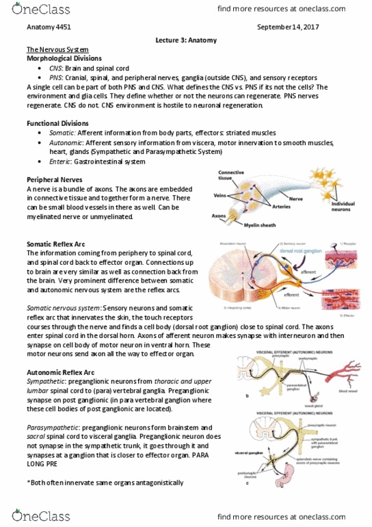
- Sensory neuron innervates the skin (touch and pain receptors)
- The axon courses through the nerve and to the DRG (where the sensory neuron cell body is)
- The DRG then connects to the spinal cord at the dorsal horn
- This afferent neuron creates a synapse in the grey matter of the spinal cord (interneuron)
- The cell body of the motor neuron is located in the ventral horn
- The motor neuron sends its axon through an efferent nerve to the striated muscles (there is no
ganglion here)
o These are the largest cells in the body – it can go from the spinal cord to a muscle in the toe
o There are morphological divisions within this single axon as it goes from the CNS (spinal cord)
to the PNS (in the muscle) in a continuous nerve
- The same CNS/PNS division can exist for the afferent nerve as it comes from the PNS (touch receptor)
to the CNS (interneuron region in spinal cord grey matter) where it forms a synapse
*At the effector
synapse, the nerve
releases ACh to the
effector muscle
find more resources at oneclass.com
find more resources at oneclass.com

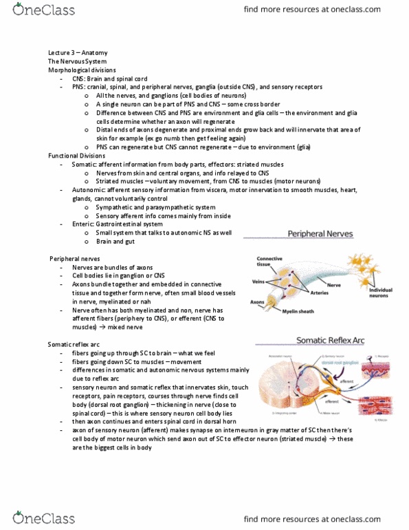
For the autonomic NS, the reflex arc differs in the effector (type of motor neurons differ in the
autonomic from the somatic). The autonomic NS also has two further divisions: sympathetic and
parasympathetic, each with their own types of reflex arcs. Both innervate the same organs, but they
have opposite effects.
1. Sympathetic (b) – fight or flight
- Preganglionic nerve that forms a synapse to a postganglionic nerve, in the para-
vertebral ganglion
- The preganglionic nerve is smaller relative to the postganglionic nerves
- This forms a sypathetic truk: the ganglions are close to the vertebra and contain all
the cell bodies of the post ganglionic nerves
- So if there is a damage to this ganglion, it can affect a whole bunch of postganglionic
nerves throughout the body
2. Parasympathetic (c) – rest and digest
- Preganglionic nerve forms a synapse to a postganglionic nerve, but not in the para-
vertebral trunk
- The preganglionic nerve is relatively longer than the postganglionic nerve
- The preganglionic nerve goes past the para-vertebral trunk and comes very close to the
effector organ, before forming the synapse to the postganglionic nerve. This synapse is
called a visceral gaglia since it is farther away from the spinal cord
- Spinal cord damage can also affect these nerves, but the effects of the damage is more
specific since there is less extensive branching of the postganglionic nerve
The sympathetic system is within the thoracic and lumbar region of the spinal cord, whereas the
parasympathetic system is located within the brainstem and sacral region of the spinal cord.
Autonomic NS releases ACh
at the preganglionic
synapse, and ACh or NE at
the effector synapse
(postganglionic to the
muscles):
- Ach = parasympathetic
- NE = sympathetic
find more resources at oneclass.com
find more resources at oneclass.com
Document Summary
Morphological divisions: cns: brain and spinal cord, pns: cranial, spinal, and peripheral nerves ganglia (outside cns), and sensory receptors. Functional divisions somatic: afferent information from body parts effectors: striated muscles (voluntary: autonomic: afferent sensory information from viscera, motor innervation to smooth muscles, heart, glands (involuntary, sympathetic and parasympathetic system, enteric: gastrointestinal system. A single neuron and some nerve bundles can be part of both the cns and the pns. It is the environment that surround those cells, not the cells themselves. Glial cells in the environment define whether an axon can regenerate or not; so whether or not they are present, determines whether the nerve/axon is part of the cns or pns, or both. Peripheral ns can regenerate and can re-innervate upon damage. However, this is not true for cells in the central ns. Both cns and pns are involved in processing both somatic and autonomic functions.