Anatomy and Cell Biology 3309 Lecture Notes - Lecture 22: Distal Convoluted Tubule, Renal Corpuscle, Proximal Tubule

22 May 2018
School
Department
Professor

Histology 3309
Urinary Passages
Learning Objectives
1. describe components, location and function of the juxtaglomerular apparatus
2. prepare a diagram of the renin-angiotensin-aldosterone system
3. compare the epithelial lining and functions of proximal tubule, loop of Henle, and distal tubule
4. describe the location and histology of the renal medulla
5. explain the structure and significance of transitional epithelium
6. outline the histological features of the urinary bladder.
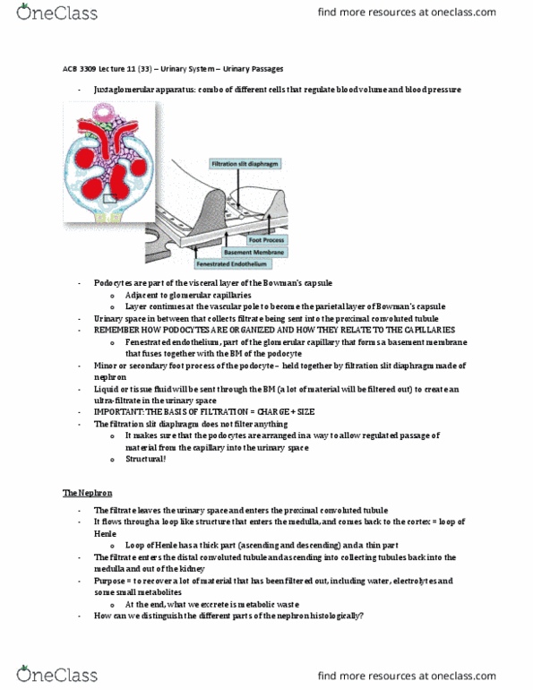
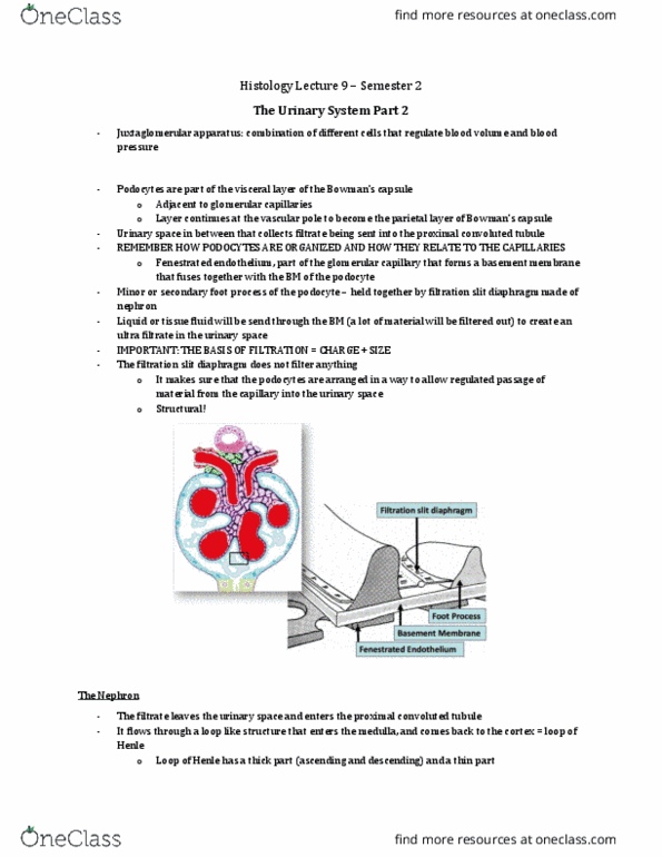
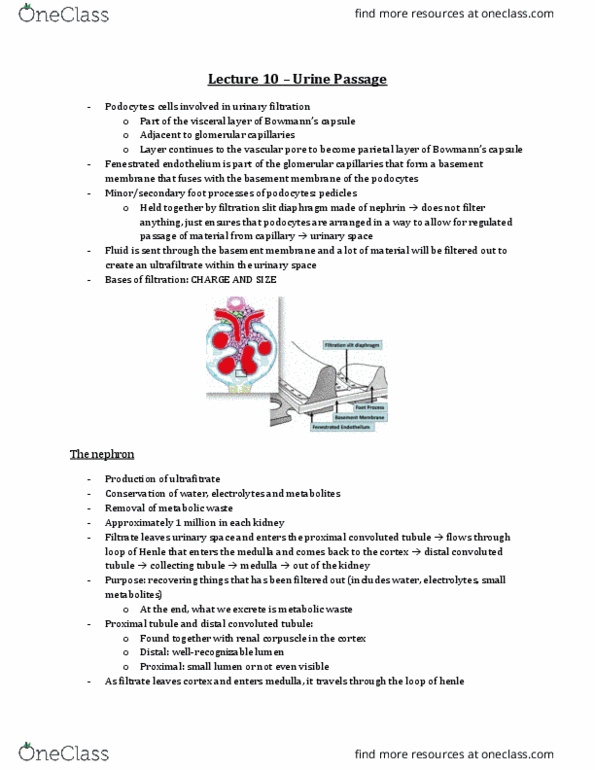
- Podocytes are part of the visceral layer of bowmans capsule adjacent to glomerular capillaries
- The layer continues around at the vascular pole to become the parietal layer of bowmans
capsule
- We also have a urinary space that collects the filtrate and sends it to the proximal convoluted
tubule
- Podocyte organization:
- We have a fenestrated capillary part of the glomerular capillary that form a BM that fuses
together w the BM of the podocyte
- This is a foot process (aka minor pedicel) (aka secondary pedicel) (aka secondary foot process)
- These are held together by filtration slit diaphragm made of nephrin
- Tissue fluid will be sent through this basement membrane and a lot of material will be filtered
out to create an ultrafiltrate within the urinary space
- Recall: basis for filtration: charge and size
The Nephron
- Production of ultrafiltrate from blood
- Conservation of water, electrolytes and metabolites
- Removal of metabolic waste
- The filtrate leaves the urinary space and enters proximal convolute tubule and then it flows the
loop of henle (that enters the medulla) and comes back up to the cortex
o Recall: the loop of henle has both a thick and a thin part
- The filtrate then enters the distal convoluted tubule and then is sent to the collecting tubule,
back into the medulla and out of the kidney
- The purpose of this is to recover a lot of material that has been filtered out (including water)
find more resources at oneclass.com
find more resources at oneclass.com

- At the end, what we excrete is just metabolic waste
- The proximal convoluted tubule + distal convoluted tubule + renal corpuscle are found in the
cortex
- Distal convoluted tubule:
o Has a well recognizable lumen
- Proximal convoluted tubule:
o Fairly small lumen (sometimes not even visible)
- Most prominent feature of the loop of henle is that part of it is thin (meaning that the epithelial
cells are squamous)
o The cells are squamous and the cytoplasm is very thin
o This allows resorption of water and NaCl through that tuble
- TS = thin segment
o Here the epithelial cells have nuclei that is very prominent but the cytoplasm is very
thin
- VR = vasa recta
o The capillaries that run parallel to the tubules to take in liquid that leaves the epithelial
lined loop of henle into those capillaries
- Collecting duct:
o Cuboidal to low columnar in shape
o Large lumen
- The proximal convoluted tubule resorbs most of the stuff (vitamins, amino acids, glucose, water,
ions, electrolytes etc)
- The filtrate then goes through the loop of henle and 2 things happen:
o The epithelum changes as it goes around the loop
o The descending limb of the loop of henle is freely permeable to water (water can easily
leave the tubule)
o This happesn bc the ascending limb of the loop of heble and the distal convoluted tubule
is IMPERMEABLE to water but it actively pumps out NaCl
o So the cells in the distal convoluted tubule and the ascending limb pump out NaCl
o This creates a high concentration of ions right outside the tubule
o SO water tries to equilibrate that and flows out at the descending limb
find more resources at oneclass.com
find more resources at oneclass.com

- 2 mechanisms for water regulation:
o aldosterone
▪ hormone
▪ produced by the adrenal gland
▪ regulates NaCl resorption from the tubule cells
▪ this indirectly regulates water reabsorption
o antidiuretic hormone (ADH) (aka vasopressin)
▪ the collecting duct has water channels within the epithelium that can be turned
on and off
▪ through the regulation of these water channels, the water reabsorption can be
regulation
▪ produced in the brain
Juxtaglomerular Apparatus
- the distal convoluted tubule originated from the renal corpuscle
- the distal convoluted tubule comes back to its own renal corpuscle at the vascular pole
- macula densa cells
o bunch of cells located right at the vascular pole
o these cells are bunched together closely (their nuclei are close together) →in light
microscopy, it shows up as a dense spot
o part of the distal convoluted tubule
o these cells measure NaCl
o they measure how efficient reabsorption of NaCl is at that nephron
- this info is conveyed to the juxtaglomerular cells
o specialized smooth muscle cells within the wall of the afferent arteriole
o these cells are both contractile and sympathetic
o JG cells produce renin
▪ Renin is a signalling molecule that starts a cascade of events
- Extraglomerular mesangial cells are also involved in the process of signal transmission bw the
macula densa and the JG cells
o There are gap junctions that are signaling things across
find more resources at oneclass.com
find more resources at oneclass.com
Document Summary
Podocytes are part of the visceral layer of bowmans capsule adjacent to glomerular capillaries. The layer continues around at the vascular pole to become the parietal layer of bowmans capsule. We also have a urinary space that collects the filtrate and sends it to the proximal convoluted tubule. We have a fenestrated capillary part of the glomerular capillary that form a bm that fuses together w the bm of the podocyte. This is a foot process (aka minor pedicel) (aka secondary pedicel) (aka secondary foot process) These are held together by filtration slit diaphragm made of nephrin. Tissue fluid will be sent through this basement membrane and a lot of material will be filtered out to create an ultrafiltrate within the urinary space. Recall: basis for filtration: charge and size. The filtrate then enters the distal convoluted tubule and then is sent to the collecting tubule, back into the medulla and out of the kidney.