Anatomy and Cell Biology 3309 Lecture Notes - Lecture 30: Dense Irregular Connective Tissue, Ovarian Follicle, Folliculogenesis

22 May 2018
School
Department
Professor

Histology 3309
Ovary and Female Reproductive System
Learning Outcomes
1. Describe the main histological features of the cortex and medulla of the ovary
2. Identify the 5 follicle types, and compare and contrast the features and cells associated with
each
3. Describe the histological changes seen during development and maturation of ovarian follicles
4. Describe the processes of ovulation and fertilization of the ovum
5. List the five hormones that regulate the female reproductive cycle and identify the cells that
produce each
6. Describe the formation of the corpus luteum and the function of its associated cells
7. Differentiate between the corpus albicans and atretic follicles
Female Reproductive System
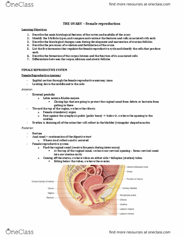
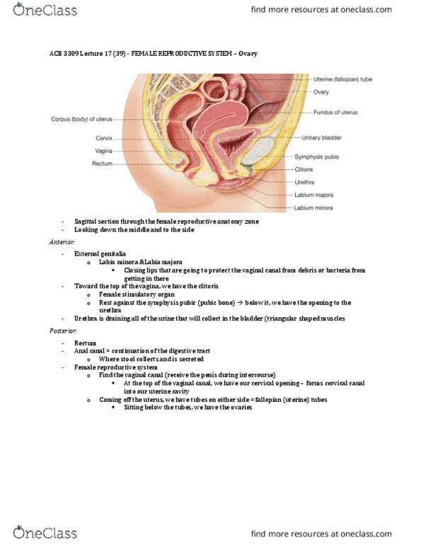
Female Reproductive Anatomy
- Labia are the closing lips that protect the vaginal canal from any debri or bacteria getting in
there
- Clitoris – female stimulatory organ – rests against the pubic bone
- Below clit is the urethra
- At the top of vaginal canal is where we have our cervical opening – forms a cervical canal into
uterine cavity
- The ovaries are anchored to the uterine body via the ovarian ligaments
- Holding them from a superior aspect is the suspensory ligament
o This ligament brings in our ovarian arteries and veins
- As the ovary goes through its cycle, it releases an egg
- It releases the egg into the environvment around it
o The ovary is located within the paritneal cavity
- This egg is released into the paritneal cavity then the fimbrae (fingerlike projections) produce
wave like motions to draw the egg into the fallopian
tube
- The egg then travels down the fallopian tube into the
fundus (top of uterus)
- If that egg is fertilizied, it will implant on the side of
the uterine cavity (in the endometrial lining)
- The endometrial lining is what thickens and falls off
during the menstrual cycle
- Myometrium is found below the endometrium
o Filled with smooth muscle
o Doesn involuntary contractions (cramping during period)
- Perimetrium: surrounding outer layer of the uterine body
- Broad ligament: drape that covers the uterus and most of the fallopian tube (Except for the
ovary and the fimbrae – these parts are projected into the peritneal cavity)
find more resources at oneclass.com
find more resources at oneclass.com

The Ovary
Ovarian General Structure
- Medulla (center)
o Where all the ovarian arteries and veins enter into
o Other things such as nerves, lymphatic vessels and loose connective tissue
- Cortex
o Where all the ovarian follicles are going through the stages of development
- Located within both the medulla and the ovarian cortex, we have an ovarian stroma (connective
tissue within the ovary)
- Sitting on top the cortex, we have a germinal epithelium
- Below that, there is a tunica albunginea (connective tissue that wraps around the ovary)
- Medulla
o can see cross sections of blood vessels, arteries and veins
- cortex:
o can see follicles at different stages in development
- zoom into the cortex
- can see ovarian follicles embedded within the ovarian stroma
- boundary of ovarian cortex:
o simple cuboidal layer of germinal epithelium
o tunica albunginea – dense irregular connective tissue
find more resources at oneclass.com
find more resources at oneclass.com

Types of Follicles
- Primordial follicles (present at birth)
- Growing follicles
o Primary follicle
▪ Unilaminar
▪ Multilaminar
o Secondary (antral) follicle
- Mature or Graafian follicles (ready for ovulation)
Clinical Case Question
- Dysgerminoma is a rare type of malignant ovarian cancer (<1%) that affects the germ cells
(oocytes) in the developing follicles. A surgeon performs a bilateral oophorectomy and sends
mboth ovaries to pathology. When examining the histopathological specimen, where would a
pathologist look to confirm this diagnosis?
a) Endometrium
b) Falloptian (uterine) tube
c) Ovarian cortex
d) Ovarine medulla
Primordial Follicle
- Present at birth
- During embryogenesis of the developing female fetus, the baby produces all her egg cells at this
time
- These eggs are stored within the ovarian cortex
- But these eggs are arrested within prophase I of meiosis I
find more resources at oneclass.com
find more resources at oneclass.com
Document Summary
Learning outcomes: describe the main histological features of the cortex and medulla of the ovary. Labia are the closing lips that protect the vaginal canal from any debri or bacteria getting in there. Clitoris female stimulatory organ rests against the pubic bone. At the top of vaginal canal is where we have our cervical opening forms a cervical canal into. Below clit is the urethra uterine cavity. The ovaries are anchored to the uterine body via the ovarian ligaments. Holding them from a superior aspect is the suspensory ligament: this ligament brings in our ovarian arteries and veins. As the ovary goes through its cycle, it releases an egg. It releases the egg into the environvment around it: the ovary is located within the paritneal cavity. This egg is released into the paritneal cavity then the fimbrae (fingerlike projections) produce wave like motions to draw the egg into the fallopian tube.