Anatomy and Cell Biology 3309 Lecture Notes - Lecture 33: Retinal Pigment Epithelium, Ciliary Body, Neural Tube

22 May 2018
School
Department
Professor

Histology 3309
Eye I
Objectives
1. Understand the organization of the eye as an organ
2. Understand the development of the eye derived from the three different germ layers
3. Understand the tissue organization of the cornea, lens, tunics, iris and ciliary body
4. Relate cell specialization to function.
5. Track the fate of light that enters the eye.
Development
- As the nervous system develops, if you have an embryo, there is a neural tube forming which
later gives rise to the spinal cord and the brain
- That neural tube is folding from the ectoderm of the embryo
- The head end of the fetus, the neural tube forms these vesicles that become the brain
- One of these vesicles then form other vesicles that later on become the retina
- They grow out of the neural tube towards the surface of the embryo at the head end
- Green = will become the retinal pigment epithelium
- Pink = neural retina (retina used for vision)
- So the vesicle grows out of the neural tube towards the surface and starts to invaginate
- At the same time, the outer surface starts to form the lens
- The ectoderm eventualy becomes the lens whereas what grew out of the neural tube becomes
like a cup
- Its still connected to the neural tube (to the rest of the brain) by this stock that will become the
optic nerve
- So these are apart of the CNS
- There is a blood brain barrier to the central nervous system and there is a blood retina barrier
to the retina
- This is a part of the brain that grew out of the brain
- The other parts of the eye are from the ectoderm (lens and the epithelium of the cornea) and
then other parts of the eye are derived from mesenchymal
find more resources at oneclass.com
find more resources at oneclass.com

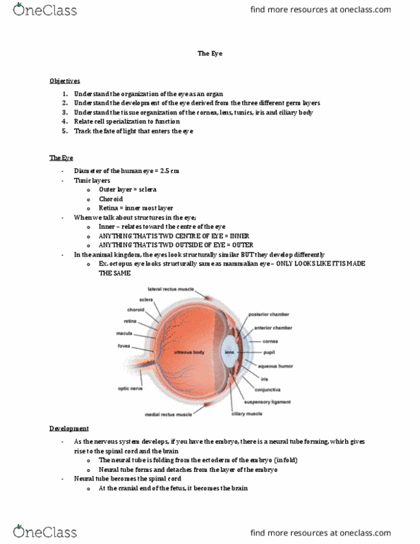
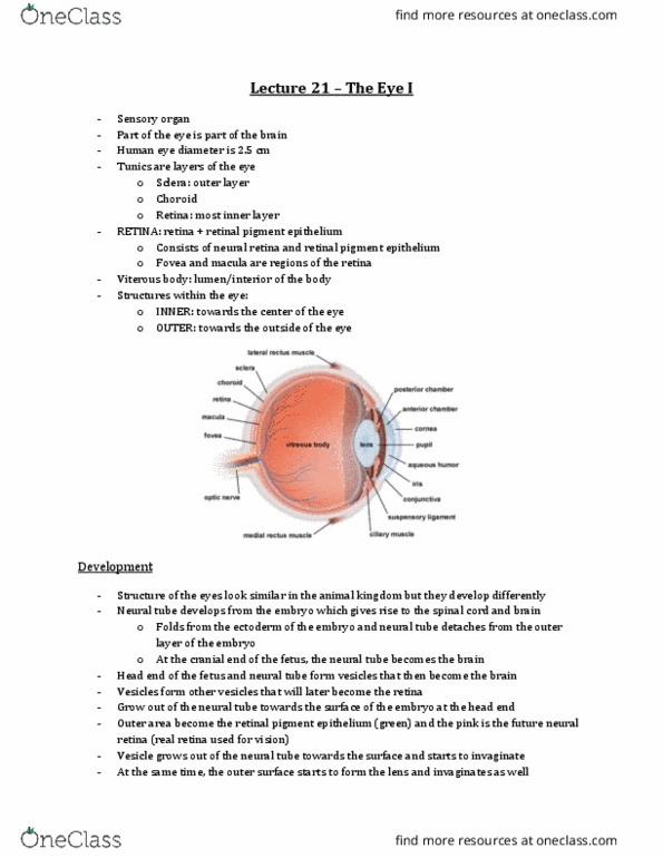
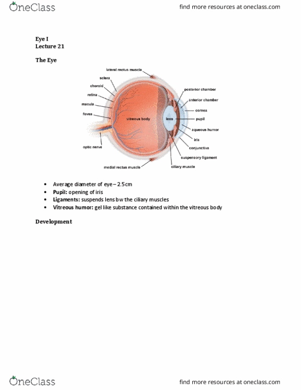
The Eye
The Tunics of the Eye
- Corneoscleral coat (cornea and sclera, ~1mm thick, protects and shapes the eye)
o Sclera is a tough layer that surrounds the eye and keeps it in shape
o Note: the sclera is continuous with the dura mater of the brain
o Sclera is also continuous with the cornea at the front of the eye
- Uvea
o Next most inner structure
o Contains…
▪ the choroid (layer that contains blood vessels)
▪ ciliary body
▪ iris
o note: the choroid is similar to the arachnoid of the brain and has the same function
- Retina
o Layer that forms out of the brain and forms the optic nerve
o Contains…
▪ Retinal Pigment Epithelium (RPE)
▪ neural retina
o retina continues as a double layer of epithelial cells at the inner edge of the ciliary body
and the iris
The Choroid
- note: sclera is the most outer part of the eye and retina is the most inner part
- the choroid is separated from the retina by bruchs membrane
- Bruch’s membrane
o Separates the retinal pigment epithelium from the choroid
o Inner most
o Basal lamina of opposing RPE and endothelial cells
find more resources at oneclass.com
find more resources at oneclass.com

- Choroid (has 2 layers):
o Choriocapillaris (inner portion)
▪ Fenestrated capillaries
▪ Supplies nutrients to the outer retina
▪ melanocytes
▪ Its very important to supply nutrients to the outer part of the retina which are
avascular so all the nutrients have to be supplied through diffusion from the
choroid which has the small capillaries
o Choroidal stroma (outer layer)
▪ Contains large vessels, nerves, collagen, fibroblasts and melanocytes
o Both layers have melanocytes bc very pigmented so if any light is not absorbed by the
retina or the retinal pigment epithelium, the choroid will make sure it doesn’t go any
further so it has dark pigmented cells
Ciliary Body
- The choroid and the sclera gives rise to the uveal portions of the eye (ciliary body and iris)
- Uveal portion
o Continuation of the choroid
o Covered by 2 layers of epithelial cells and they secrete the aqueous humor but they also
important bc they have fibers inserted in them which are attached to the lens
o Contains ciliary muscles (radial & circular) and if these muscles constrict, it will stretch
the lens so the opening gets smaller and the fibers connected to the ciliar body open up
and so our lens can round up →this is how we accommodate to something that is close
by
o When we looked into something that is far away, our ciliary muscle is relaxed and our
fibers are stretched flat so the lens is stretched as well →this is how we accommodate to
see things that are far
o As you age, you cant really see things that are close anymore bc once your ciliary muscle
constricts and your fibers loosen up, your lens doesn’t round up as much as it used to bc
it loses elasticity
o contains fenestrated capillaries
o Accommodation!
- Note: there are 2 layers of epithelial covering the ciliary body
o These layres are continuous with the retinal pigment epithelium and the neuralretina
- Neuroepithelial portion (continuation with retina)
o Contributes the two layers of the ciliary epithelium
find more resources at oneclass.com
find more resources at oneclass.com
Document Summary
As the nervous system develops, if you have an embryo, there is a neural tube forming which later gives rise to the spinal cord and the brain. That neural tube is folding from the ectoderm of the embryo. The head end of the fetus, the neural tube forms these vesicles that become the brain. One of these vesicles then form other vesicles that later on become the retina. They grow out of the neural tube towards the surface of the embryo at the head end. Green = will become the retinal pigment epithelium. Pink = neural retina (retina used for vision) At the same time, the outer surface starts to form the lens. The ectoderm eventualy becomes the lens whereas what grew out of the neural tube becomes. So the vesicle grows out of the neural tube towards the surface and starts to invaginate like a cup.