Anatomy and Cell Biology 3309 Lecture Notes - Lecture 35: Ear Canal, Oval Window, Earwax

22 May 2018
School
Department
Professor

Histology 3309
The Ear
Objectives
- Describe the anatomy of the ear
- Describe the organization of the middle ear
- Describe the organization of the cochlea
- Describe how the organ of Corti is designed to translate mechanic movement into electrical cell
signals
- Explain how the hair cells function
- Explain the way sound is transduced into mechanical movement in the middle ear, and into cell
signals in the cochlea
Anatomy of the Ear
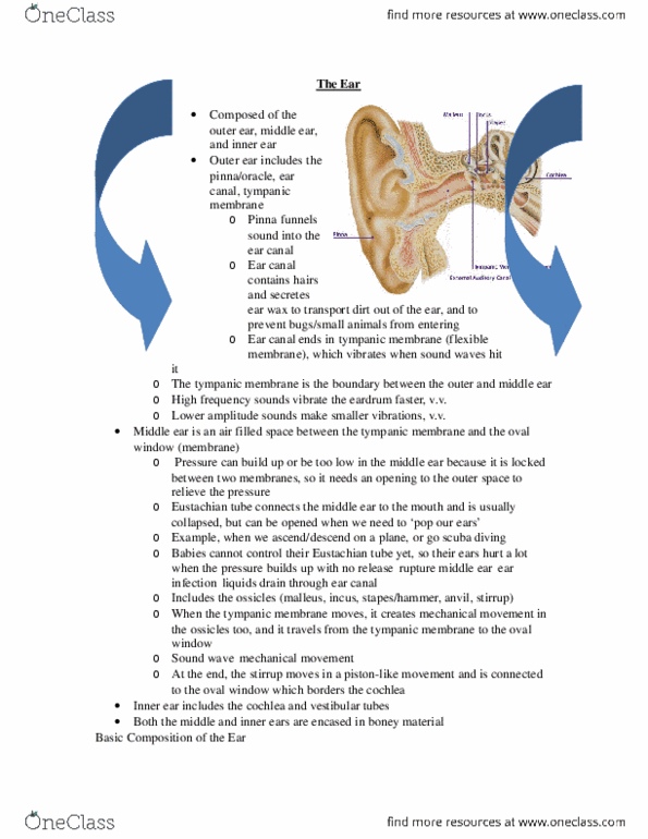
- Pinna: outside of ear (mostly cartilage tissue)
o Funnels the soundwaves into the external auditory ear canal
- The outer ear canal contains glands that secrete ear wax
o Ear wax keeps the ear canal open and clean
- There are hairs in the external auditory canal that prevents the insects from entering the canal
and help expel any dirt from the inside of the external auditory canal
- The external auditory canal ends with the eardrum (aka tympanic membrane)
- When sound waves enter the ear canal, they will hit on the tympanic membrane, which will
move the ear drum
- The ear drum is also the border bw the external ear and the middle ear
- The middle ear is a cavity behind the ear drum – its an air filled space that ends with the oval
window
- This contains the ossicles (3 little bones – malleus, incus and stapes)
o Note: latin word for stapes is stirrup
- The malleus picks up the movement of the eardrum and it conveys the movement to the incus
which is linked to the stapes
- So the ossicles pick up the movement of the eardrum and amplify it and convert it into a
movement against the oval window
- Air can expand depending on the pressure and temperature
- So the air filled space of the middle ear has to somehow be connected to the exterior bc
otherwise we get high or low pressure in the middle ear
- In order to have the high or low pressure adapted to the exterior air pressure, the middle ear is
connected to the nasal pharaynx (mouth) by the Eustachian tube
o This tube is normally collapsed
but when you crack youre ear, it
opens and you release the air
pressure in your middle ear
- Cochlea is what we use to hear
o It is innervated by the auditory
and vestibular nerves (joint
nerves)
o So it’s the 8th nerve that
innervates both the vestibular
and auditory system
find more resources at oneclass.com
find more resources at oneclass.com

Organization of the Auditory System
- sound hits the ear drum, it makes the ossicles (stirrup) move against the oval window
(membrane that separates the cochlea from the middle ear)
- the cochlea has a space filled with perilymph liquid
- the oval window moves against the liquid
- we cant compress liquids so the only way the oval window can move against the perilymph is at
the other end there is a round window (another membrane)
- as the stirrup presses against the oval window, the entire liquid moves to the apex and back on
the other side through the perilymph space against the round window
- when sound enters our ear, the stirrup presses against the perilymph space and the liquid starts
to move all the way up and down the coil
- the perilymph space also has another fluid filled space filled with endolymph (high K+ solution)
- within the endolymph space, we have the organ of corti
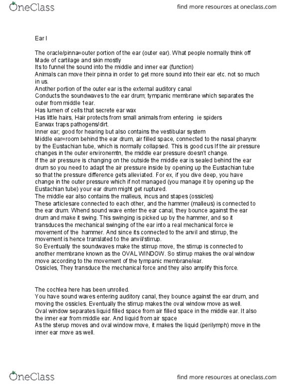
Organization of the Auditory System
- m=malleus
- i= incus
- s= stapes
- stapes moves against the perilymph space all the way to the tip (center of the coil) and then all
the way back to the round window
- the endolymph filled space is embedded within that
- the endolymph filled space contains a membrane called the basilar membrane
o the basilar membrane has specific structural traits
o its stiff at the beginning
o as it extends to the helicotrema (tip of the coil), it becomes more floppy
o so depending on which frequency the stirrup pounds against the oval window, the
basilar membrane starts to swing
o so high frequencies (ex: 20kHz) make the basilar membrane swing a lot at the beginning
o if you have low frequencies (ex: 0.2 Hz), the basilar membrane swings near the tip
- so diff sound frequencies are distributed along the basilar membrane according to where it
swings optimally in response to a certain sound frequency
find more resources at oneclass.com
find more resources at oneclass.com
Document Summary
Describe the organization of the middle ear. Describe how the organ of corti is designed to translate mechanic movement into electrical cell signals. Explain the way sound is transduced into mechanical movement in the middle ear, and into cell signals in the cochlea. Pinna: outside of ear (mostly cartilage tissue: funnels the soundwaves into the external auditory ear canal. The outer ear canal contains glands that secrete ear wax: ear wax keeps the ear canal open and clean. There are hairs in the external auditory canal that prevents the insects from entering the canal and help expel any dirt from the inside of the external auditory canal. The external auditory canal ends with the eardrum (aka tympanic membrane) When sound waves enter the ear canal, they will hit on the tympanic membrane, which will move the ear drum. The ear drum is also the border bw the external ear and the middle ear.