Anatomy and Cell Biology 3319 Lecture Notes - Lecture 39: Common Hepatic Duct, Common Bile Duct, Mesentery

1 May 2018
School
Department
Professor

Lecture 039: Small Intestine, Large Intestine, and Accessory Organs
Objectives
● Describe the duodenum and its associated structures (ie pancreas and gallbladder).
● Describe the visceral surface of the liver. What structures entering the porta hepatis
contribute to the “triads” of the liver ?
● Describe the distinguishing features of the jejunum and ileum
● Define the following terms: epiploic appendages, plicae semilunares, taenia coli and
haustra.
● Describe the anal canal and the processes involved with defecation
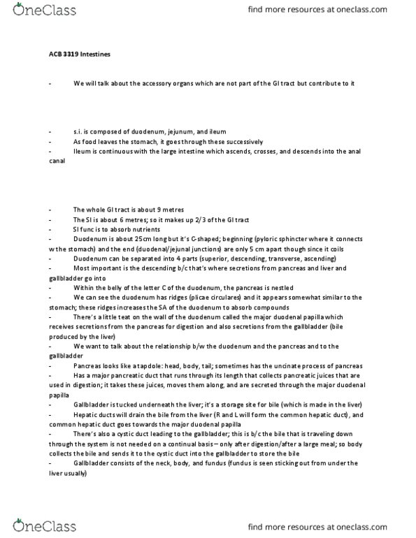
The small intestine
● Total length: 6 m
● Divided into 3 sections
○ Duodenum
■ 25 cm
○ Jejunum
■ About of the small intestine⅖
○ Ileum
■ About of the small intestine⅗
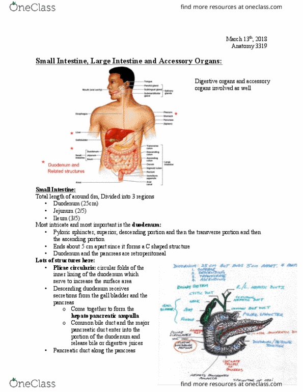
Duodenum
● Ends 5 cm apart because it is a C-shaped structure
● Made of 4 sections:
○ Superior, descending, transverse, ascending
● Is a retroperitoneal structure
○ Dives behind the peritoneum and emerges later at the end
● Plicae circulares
○ Circular folds of the inner lining
○ Increases the surface area of the duodenum
● Descending dodum is the most important section
○ Receives secretions from the pancreas and gallbladder
■ Via the hepatopancreatic ampulla (a merging of the common bile duct
and the major pancreatic duct)
■ Gets bile or digestive juices
Duodenum, pancreas and biliary system
● Bile is made in the liver
○ Collected by the right and left hepatic duct
■ Merges into the common hepatic duct
○ Travels to duodenum via the hepatopancreatic ampulla
○ Or stored in the gallbladder via the cystic duct
●Gallbladder
find more resources at oneclass.com
find more resources at oneclass.com

○ Stores and concentrate
bile
● Duodenum releases
cholecystokinin (CCK) in
response to a fatty meal
○ CCK travels in circulation
to the gallbladder
■ Causes muscular
wall contraction
■ Force bile release
along the cystic
duct to the bile duct
●Gallstones
○ Due to precipitation of bile
salts
○ Can cause pain in the
gallbladder when it contracts
○ Can become lodged in the biliary system
■ Can block the release of stored bile if it gets lodged in the cystic duct
■ Can block all release of bile (stored and fresh) if it gets lodged in the bile
duct
The Pancreas
● A secondarily retroperitoneal structure
○ Is lodged behind the c-shape of the duodenum
● Both an endocrine and an exocrine organ,
○ Many exocrine acinar glands
○ Fewer clusters of endocrine cells
● Exocrine acinar cells
○ Secrete 22 kinds of pancreatic enzymes for digestion
○ Enzymes (proteases) are stored within the cells as inactive zymogen granules
■ Become activated in the duodenum
○ Collected through the small ducts that lead into the hepatopancreatic ampulla
● Islets of Langerhans (pancreatic islets)
○ Carry out the endocrine function
○ Secretes insulin (� cells)
○ Secretes glucagon (α cells)
○
Functions of the Liver
● The liver has more than 200 important functions
● Synthesis and secretion of bile
● Storage of glycogen and lipid reserves
● Synthesis and release of cholesterol bound to transport proteins
find more resources at oneclass.com
find more resources at oneclass.com

● Inactivation of toxins
○ Toxic compounds (alcohol, carbon tetrachloride) are carried to the liver via
hepatic portal vein and are metabolically inactive
● Maintenance of normal blood glucose, amino acid and fatty acid concentrations
● Blood storage (major contributor to venous reserve)
● Synthesis of plasma proteins and clotting factors
Mesenteries
● Double folds of peritoneum (joins the visceral and parietal layers)
●Greater momentum
○ Important site for fat storage
○ Attaches to the transverse colon
●Transverse mesocolon
●Sigmoid mesocolon
Mesenteries of the liver
●Falciform ligament
○ Attaches the liver to the anterior abdominal wall
●Lesser omentum
○ Attaches the liver to the
stomach
Liver
● Anterior (diaphragmatic) surface
○ Tucked underneath the
diaphragm
○ Falciform ligament
■ Divides the liver into
the left and right lobe
● Visceral surface of the liver
○Porta hepatis
■ Everything
traveling to and
away from the
liver must pass
through this
opening
● Hepatic
portal vein
● Hepatic
artery
● Hepatic
duct
■ VERY
find more resources at oneclass.com
find more resources at oneclass.com
Document Summary
Lecture 039: small intestine, large intestine, and accessory organs. Describe the duodenum and its associated structures (ie pancreas and gallbladder). Describe the visceral surface of the liver. Describe the distinguishing features of the jejunum and ileum. Define the following terms: epiploic appendages, plicae semilunares, taenia coli and haustra. Describe the anal canal and the processes involved with defecation. Ends 5 cm apart because it is a c-shaped structure. Dives behind the peritoneum and emerges later at the end. Increases the surface area of the duodenum. Descending dodum is the most important section. Receives secretions from the pancreas and gallbladder. Via the hepatopancreatic ampulla (a merging of the common bile duct and the major pancreatic duct) Collected by the right and left hepatic duct. Travels to duodenum via the hepatopancreatic ampulla. Or stored in the gallbladder via the cystic duct. Duodenum releases cholecystokinin (cck) in response to a fatty meal. Cck travels in circulation to the gallbladder.